Japanese
English
- 有料閲覧
PPV購入案内
1,320円 (1,200円+税10%) こちらは、1465ページから1469ページの文献です。購入には医書.jp本会員登録が必要です。
会員登録完了後に改めて上記「購入サイトへ」を選択してください。
または「購入サイトへ」選択後に「購入ログイン」、「新規会員登録」の順に進んでください。
会員登録について詳しくはをご確認ください。
- Abstract 文献概要
- 1ページ目 Look Inside
- 参考文献 Reference
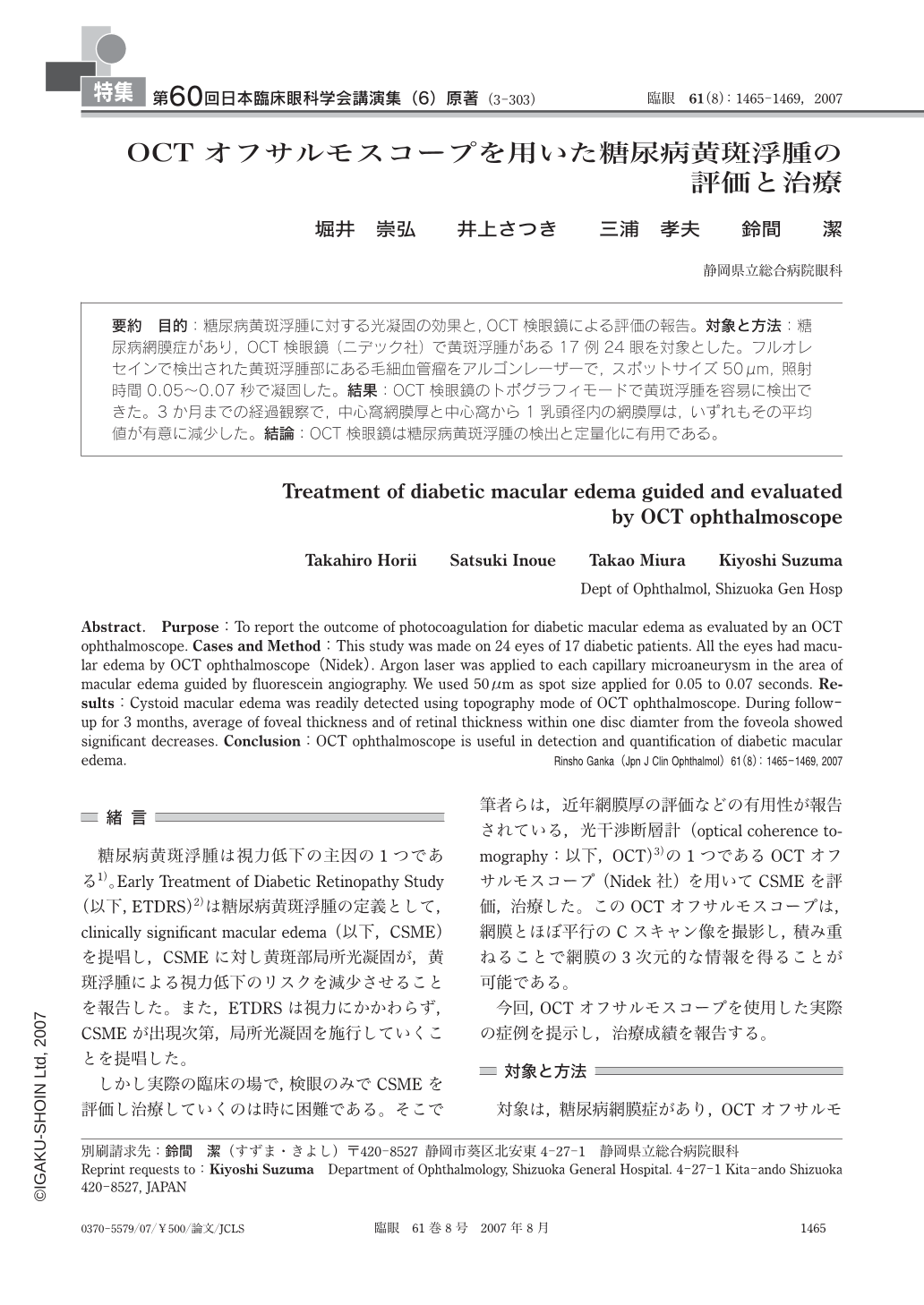
要約 目的:糖尿病黄斑浮腫に対する光凝固の効果と,OCT検眼鏡による評価の報告。対象と方法:糖尿病網膜症があり,OCT検眼鏡(ニデック社)で黄斑浮腫がある17例24眼を対象とした。フルオレセインで検出された黄斑浮腫部にある毛細血管瘤をアルゴンレーザーで,スポットサイズ50μm,照射時間0.05~0.07秒で凝固した。結果:OCT検眼鏡のトポグラフィモードで黄斑浮腫を容易に検出できた。3か月までの経過観察で,中心窩網膜厚と中心窩から1乳頭径内の網膜厚は,いずれもその平均値が有意に減少した。結論:OCT検眼鏡は糖尿病黄斑浮腫の検出と定量化に有用である。
Abstract. Purpose:To report the outcome of photocoagulation for diabetic macular edema as evaluated by an OCT ophthalmoscope. Cases and Method:This study was made on 24 eyes of 17 diabetic patients. All the eyes had macular edema by OCT ophthalmoscope(Nidek). Argon laser was applied to each capillary microaneurysm in the area of macular edema guided by fluorescein angiography. We used 50μm as spot size applied for 0.05 to 0.07 seconds. Results:Cystoid macular edema was readily detected using topography mode of OCT ophthalmoscope. During follow-up for 3 months, average of foveal thickness and of retinal thickness within one disc diamter from the foveola showed significant decreases. Conclusion:OCT ophthalmoscope is useful in detection and quantification of diabetic macular edema.
Copyright © 2007, Igaku-Shoin Ltd. All rights reserved.