Japanese
English
- 有料閲覧
PPV購入案内
1,320円 (1,200円+税10%) こちらは、955ページから958ページの文献です。購入には医書.jp本会員登録が必要です。
会員登録完了後に改めて上記「購入サイトへ」を選択してください。
または「購入サイトへ」選択後に「購入ログイン」、「新規会員登録」の順に進んでください。
会員登録について詳しくはをご確認ください。
- Abstract 文献概要
- 1ページ目 Look Inside
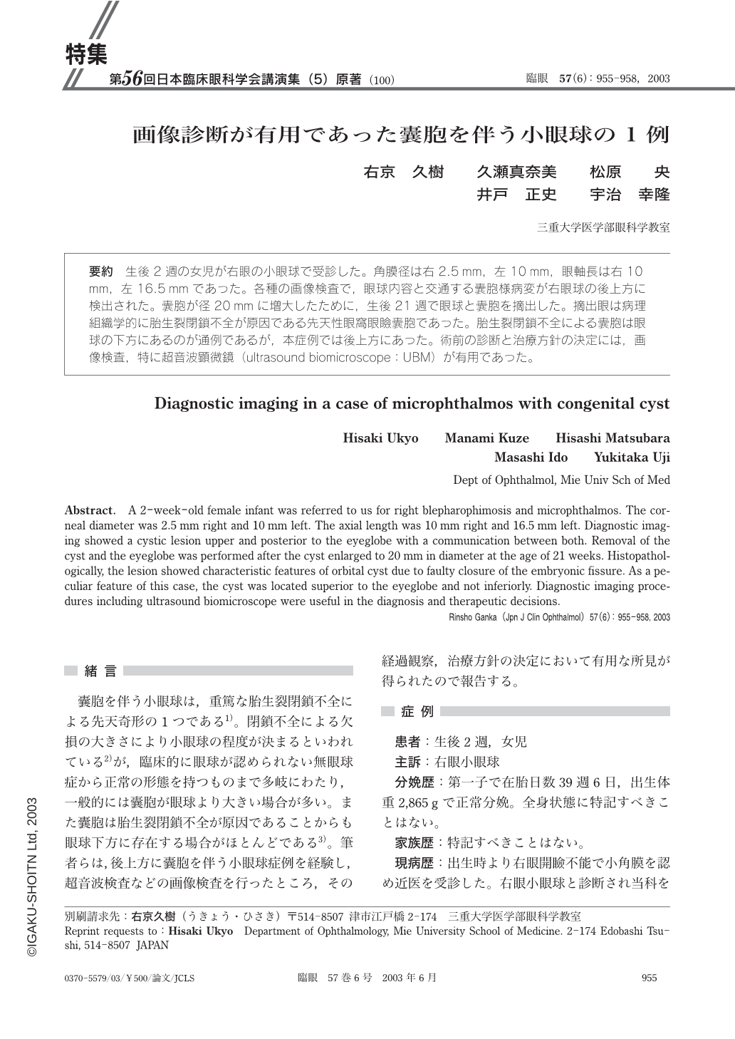
要約 生後2週の女児が右眼の小眼球で受診した。角膜径は右2.5mm,左10mm,眼軸長は右10mm,左16.5mmであった。各種の画像検査で,眼球内容と交通する囊胞様病変が右眼球の後上方に検出された。囊胞が径20mmに増大したために,生後21週で眼球と囊胞を摘出した。摘出眼は病理組織学的に胎生裂閉鎖不全が原因である先天性眼窩眼瞼囊胞であった。胎生裂閉鎖不全による囊胞は眼球の下方にあるのが通例であるが,本症例では後上方にあった。術前の診断と治療方針の決定には,画像検査,特に超音波顕微鏡(ultrasound biomicroscope:UBM)が有用であった。
Abstract. A 2-week-old female infant was referred to us for right blepharophimosis and microphthalmos. The corneal diameter was 2.5 mm right and 10 mm left. The axial length was 10 mm right and 16.5 mm left. Diagnostic imaging showed a cystic lesion upper and posterior to the eyeglobe with a communication between both. Removal of the cyst and the eyeglobe was performed after the cyst enlarged to 20 mm in diameter at the age of 21 weeks. Histopathologically,the lesion showed characteristic features of orbital cyst due to faulty closure of the embryonic fissure. As a peculiar feature of this case,the cyst was located superior to the eyeglobe and not inferiorly. Diagnostic imaging procedures including ultrasound biomicroscope were useful in the diagnosis and therapeutic decisions.
Copyright © 2003, Igaku-Shoin Ltd. All rights reserved.