Japanese
English
- 有料閲覧
PPV購入案内
1,320円 (1,200円+税10%) こちらは、533ページから538ページの文献です。購入には医書.jp本会員登録が必要です。
会員登録完了後に改めて上記「購入サイトへ」を選択してください。
または「購入サイトへ」選択後に「購入ログイン」、「新規会員登録」の順に進んでください。
会員登録について詳しくはをご確認ください。
- Abstract 文献概要
- 1ページ目 Look Inside
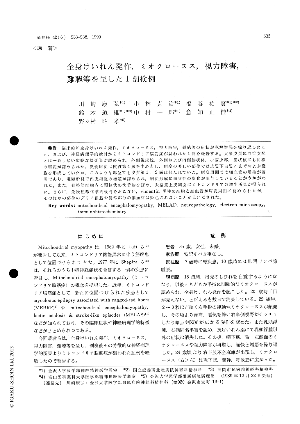
臨床的に全身けいれん発作,ミオクローヌス,視力障害,難聴等の症状が寛解増悪を繰り返したこと,および,神経病理学的検討からミトコンドリア脳筋症が疑われた1例を報告する。大脳皮質に血管支配とは一致しない広範な壊死巣が認められ,外側視床枕,外側および内側膝状体,小脳虫部,歯状核にも同様の病変が認められた。皮質病変は皮質第4層を中心とし,病変の著しい部位では皮質下白質にまでおよぶ嚢胞を形成していたが,このような部位でも皮質第1,2層は保たれていた。病変周囲では細血管の増生が著明であり,電顕所見で内皮細胞の増殖が認められ,病変形成に血管性の変化が関与していることがうかがわれた。また,骨格筋細胞内に穎粒状の沈着物を認め,脈絡叢上皮細胞にミトコンドリアの増生所見が得られた。さらに,免疫組織化学的検討をおこない,vimentin陽性の細胞と細血管が病変周囲に認められたが,そのほかの部位のグリア細胞や健常部分の細血管は染色されないことが見いだされた。
We have reported the clinical and autopsy findings in a case with generalized seizures, myoclonus, blindness and deafness which was accompanied by stroke-like episodes. This case was diagnosed as mitochondrial encephalomyopa-thy, lactic acidosis & stroke-like episodes (MELAS) from these findings. Solitary and continuous lesions of softening were distributed in both hemispheres, more severely in the frontal and occipital poles. These lesions did not correspond to a vascular supply. The pulvinar, lateral and medial geniculate body of the thalamus, cerebellar vermis and dentate nucleus had small lesions of softening. The cortical lesions occurred mainly in layer 4, and the most prominent lesions among them appeared cystic, involving the subcortical white matter, but nerve cells in layer 1 and 2 were preserved. Proliferation of small blood vessels was seen around the softening areas. Electron microscopy revealed increased mitochondria in endothelial cells of these vessels, abnormal dense bodies in skeletal muscle cells and tightly packed mitochondria in choroid plexus epithelial cells. Immunohistochemical study suggested that vimen-tin positive cells were seen around lesions and proliferated vessels are different from those seen in the intact tissues.
Copyright © 1990, Igaku-Shoin Ltd. All rights reserved.