Japanese
English
- 有料閲覧
PPV購入案内
1,320円 (1,200円+税10%) こちらは、815ページから824ページの文献です。購入には医書.jp本会員登録が必要です。
会員登録完了後に改めて上記「購入サイトへ」を選択してください。
または「購入サイトへ」選択後に「購入ログイン」、「新規会員登録」の順に進んでください。
会員登録について詳しくはをご確認ください。
- Abstract 文献概要
- 1ページ目 Look Inside
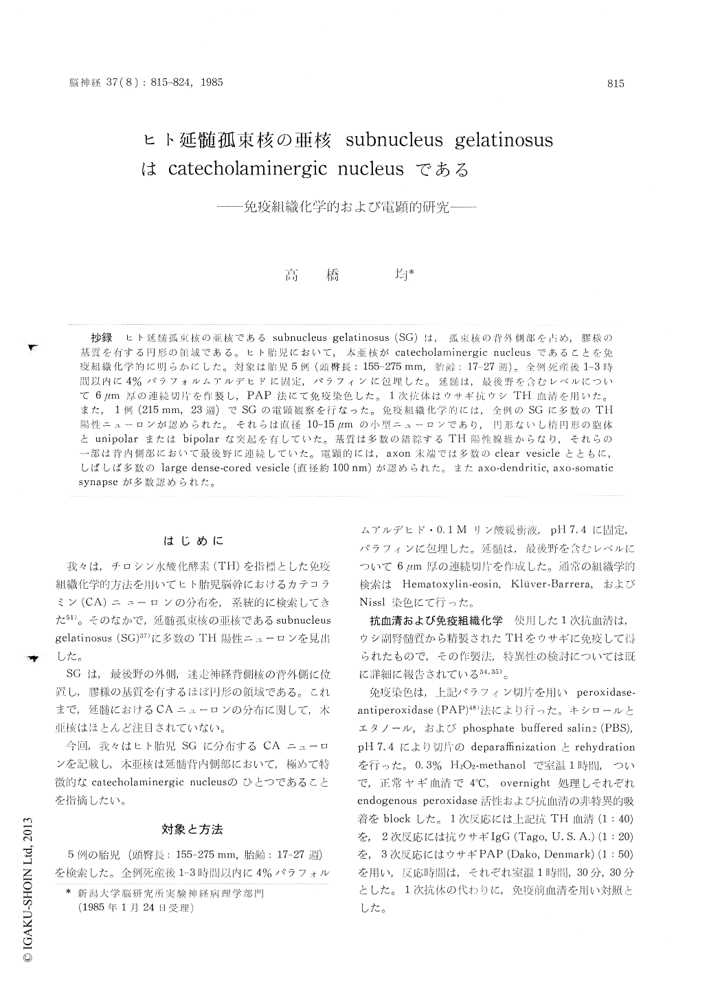
抄録 ヒト延髄孤束核の亜核であるsubnucleus gelatinosus (SG)は,孤束核の背外側部を占め,膠様の基質を有する円形の領域である。ヒト胎児において,本亜核がcatecholamincrgic nucleusであることを免疫紅織化学的に明らかにした。対象は胎児5例(頭臀長;155-275mm,胎齢:17-27週)。全例死産後1-3時間以内に4%パラフォルムアルデヒドに固定,パラフィンに包埋した。延髄は,最後野を含むレベルについて6μm厚の連続切片を作製し,PAP法にて免疫染色した。1次抗体はウサギ抗ウシTH血清を用いた。また,1例(215mm,23週)でSGの電顕観察を行なった。免疫組織化学的には,全例のSGに多数のTH陽性ニューロンが認められた。それらは直径10-15μmの小型ニューロンであり,円形ないし楕円形の胞体とunipolarまたはbipolarな突起を有していた。基質は多数の錯綜するTH陽性線維からなり,それらの一部は背内側部において最後野に連続していた。電顕的には,axon末端では多数のclear vesicleとともに,しばしば多数のlarge dense-cored vesicle (直径約100nm)が認められた。またaxo-dendritic,axo-somaticsynapseが多数認められた。
The subnucleus gelatinosus of the nucleus tractus solitarii (SG) has been described in man by Olszewski and Baxter (1954). It is located in the dorsolateral corner of the nucleus tractus solitarii at the level of the area postrema and appears as the small, round area characterized by a peculiar gelatinous appearance of the ground substance. In this communication, we describe the existence of many catecholamine neurons in this particular area of human fetuses.
We examined the lower medulla oblongata of five human fetuses (CRL: 155-275 mm, GA: 17-27 wks).
The brains were obtained within 1-3 hours after death following therapeutic or spontaneous abortions. They were immedately fixed with 4% paraformaldehyde in 0.1 M phosphate buffer, pH 7.4, dehydrated graded alcohol, and embedded in paraffin. Serial 6 μm-sections containing the SG were cut from the lower medulla oblongata of each fetus. These sections were stained by pero-xidase-antiperoxidase (PAP) technique using rabbit anti-bovine TH sera. The preparation and the specificity of the antisera used were described elsewhere (Nakashima et al, 1983 & 1984). In addition, ultrastructural examination was made on the unilateral SG of one fetus (CRL: 215 mm, GA: 23 wks).
Immunohistochemically, many TH-positive neur-ons were observed in the SG of all cases. They were fairly uniform in size, 10-15 pm in diameter, showing round to oval cell somata and unipolar or bipolar processes. In the neuropil, numerous TH-positive fibers were found oriented randomly. Some of TH-positive fibers were seen extending toward the area postrema in the dorsomedial region of this subnucleus. A small number of TH-negative neurons were also noted.
Ultrastructurally, the neurons observed were small in size and possessed in general a scanty cytoplasmic rim around the round nucleus. In the neuropil, unmyelinated nerve fibers, axon terminals with synaptic vesicles, dendrites and glial cell processes were observed. Synaptic structures encountered were axo-dendritic and axo-somatic. However, axo-axonic synapses could not be found. It was of interest that large dense-cored vesicles were frequently seen together with clear vesicles in the axon terminals.
There have been so far many reports with regard to the distribution of the catecholamine neurons in the central nervous system of various mammals including man. In the medulla oblongata, the dorsomedial catecholamine neurons have been well known as the A2 group since the first des.cription in rats by DahlstrOm and Fuxe (1964).Onthe anatomical basis, however, little attention hasbeen paid to the SG and the equivalent area inexperimental mammals.In the present immuno-histochemical study made on the human fetuses,it was clearly demonstrated that the SG containsmany catecholamine neurons, indicating that itis a catecholaminergic nucleus.
Copyright © 1985, Igaku-Shoin Ltd. All rights reserved.