Japanese
English
- 有料閲覧
PPV購入案内
1,320円 (1,200円+税10%) こちらは、781ページから796ページの文献です。購入には医書.jp本会員登録が必要です。
会員登録完了後に改めて上記「購入サイトへ」を選択してください。
または「購入サイトへ」選択後に「購入ログイン」、「新規会員登録」の順に進んでください。
会員登録について詳しくはをご確認ください。
- Abstract 文献概要
- 1ページ目 Look Inside
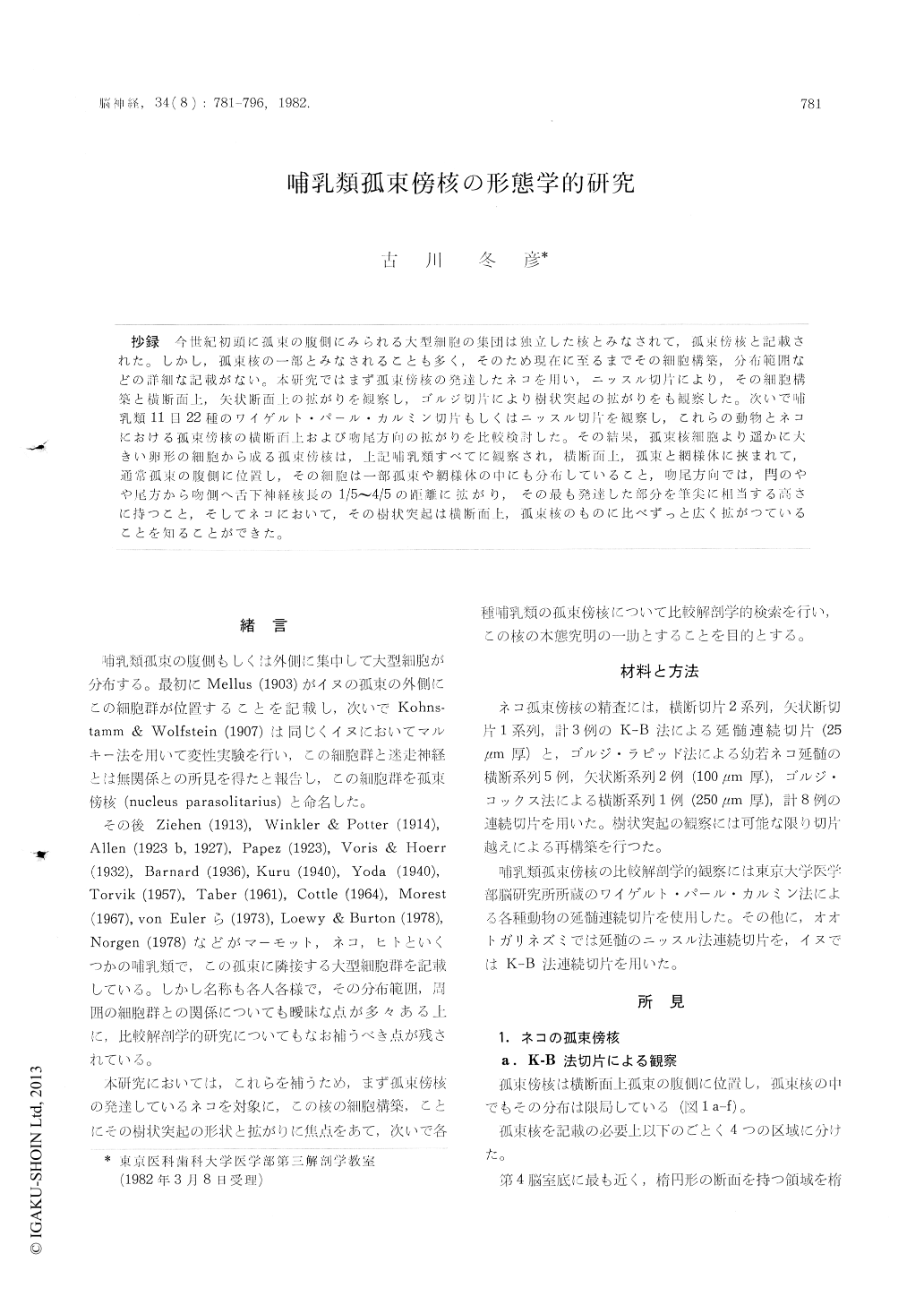
抄録 今世紀初頭に孤束の腹側にみられる大型細胞の集団は独立した核とみなされて,孤束傍核と記載された。しかし,孤束核の一部とみなされることも多く,そのため現在に至るまでその細胞構築,分布範囲などの詳細な記載がない。本研究ではまず孤束傍核の発達したネコを用い,ニッスル切片により,その細胞構築と横断面上,矢状断面上の拡がりを観察し,ゴルジ切片により樹状突起の拡がりをも観察した。次いで哺乳類11目22種のワイゲルト・パール・カルミン切片もしくはニッスル切片を観察し,これらの動物とネコにおける孤束傍核の横断面上および吻尾方向の拡がりを比較検討した。その結果,孤束核細胞より遥かに大きい卵形の細胞から成る孤束傍核は,上記哺乳類すべてに観察され,横断面上,孤束と網様体に挾まれて,通常孤束の腹側に位置し,その細胞は一部孤束や網様体の中にも分布していること,吻尾方向では,閂のやや尾方から吻側へ舌下神経核長の1/5〜4/5の距離に拡がり,その最も発達した部分を筆尖に相当する高さに持つこと,そしてネコにおいて,その樹状突起は横断面上,孤束核のものに比べずっと広く拡がつていることを知ることができた。
A comparative anatomical study of the parasoli-tary nucleus (Kohnstamm & Wolfstein, 1907) using Weigert-Pal-Carmine or Nissle sections of mammalian brain-stem was made to discover its precise topography and cytoarchitecture.
In transverse planes, the nucleus is usually found on the ventral side of the solitary tract. The parasolitary cells can be found not only among the solitary cells, but in the solitary tract, and in the parvocellular reticular formation (Fig. 1 a-f & 4). The oval or drop shaped parasolitary cells can be easily identified because they are nearly as large as the hypoglossal cells (Table 1). The cells of the solitary nucleus and parvocellular reticular formation are only one half to two thirds as large as the parasolitary cells.
In sagittal planes, the mammalian parasolitary nucleus is one to four fifths as long as the hypo-glossal nucleus. Its most developed part can be found somewhat oral to the obex (Fig. 1 h & 5).
A Golgi study of the cat parasolitary nucleus was also made, using Golgi-Rapid and Golgi-Cox serial sections. Three-dimentional reconstruction with serial sections was attempted as far as possible.
The dendrites of the cat parasolitary cells are thick and straight with blunted tips. They show only a few ramifications. The longest dendrite extends as far as 700 pm. The directions of the dendrites are variable. Each cell has eight to ten dendrites. Few spines can be observed (Fig. 2 a,b).
The dendritic field of the parasolitary nucleus, as a whole, extends dorsally into the middle layer of the solitary nucleus, ventrally into the parvocellular reticular formation, medially into the intercalate nucleus, and laterally into the inferior nucleus of the vestibular nerve (Fig. 3).
Copyright © 1982, Igaku-Shoin Ltd. All rights reserved.