Japanese
English
- 有料閲覧
- Abstract 文献概要
- 1ページ目 Look Inside
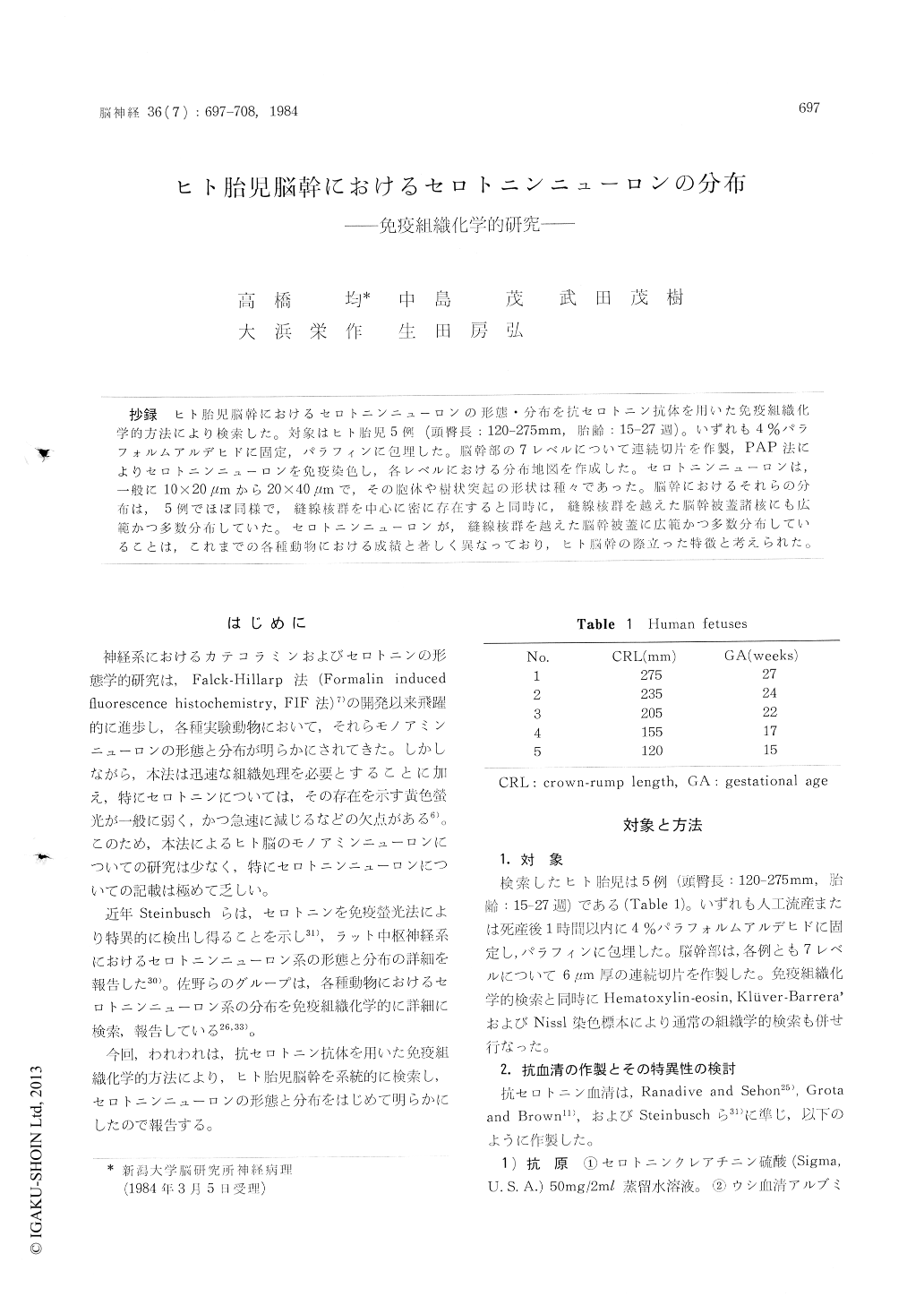
抄録 ヒト胎児脳幹におけるセロトニンニューロンの形態・分布を抗セロトニン抗体を用いた免疫組織化学的方法により検索した。対象はヒト胎児5例(頭臀長:120-275mm,胎齢:15-27週)。いずれも4%パラフォルムアルデヒドに固定,パラフィンに包埋した。脳幹部の7レベルについて連続切片を作製,PAP法によりセロトニンニューロンを免疫染色し,各レベルにおける分布地図を作成した。セロトニンニューロンは,一般に10×20μmから20×40μmで,その胞体や樹状突起の形状は種々であった。脳幹におけるそれらの分布は,5例でほぼ同様で,縫線核群を中心に密に存在すると同時に,縫線核群を越えた脳幹被蓋諸核にも広範かつ多数分布していた。セロトニンニューロンが,縫線核群を越えた脳幹被蓋に広範かつ多数分布していることは,これまでの各種動物における成績と著しく異なっており,ヒト脳幹の際立った特徴と考えられた。
The distribution of serotonin neurons in the central nervous system (CNS) has been intensively examined in mammals, such as rats, cats and monkeys. However, the details of serotonin neuron system have been remained uncertain in human CNS, although two fluorescence histochemical studies were reported in human fetus.
In this study, we performed immunohistochemical examination on the distribution of serotonin neurons in the brain stem of human fetuses.
The brain stems from five human fetuses (CRI, : 120-275 mm, GA : 15-27 wks) were fixed with 4% paraformaldehyde, dehydrated with graded ethanol, and embedded in paraffin. Serial sections, 6 inn in thickness, were cut from seven different levels of the brain stem of each fetus. The initial several sections were used for usual histological observations. The following serial ones were stained by peroxidase-antiperoxidase (PAP) tech-nique using anti-serotonin sera. The anti-serotonin sera used were raised in rabbits by the methcds of Ranadive and Sehon (1967), Grota and Brown (1974), and Steinbusch et al (1978). Before the use, the specificity of the antisera was confirmed by the immunohistochemical examination of the CNS of rat embryos and adults.
Positively stained serotonin neurons were clearly demonstrated in the brain stems of all cases examined (Figs. 1A-1H). They were small to medium in size, 10 x20 /tin to 20 x40 pm, and varied in shape, showing round to oval cell scmata with unipolar, bipolar and multipolar processes. The distribution of serotonin neurons in the brain stems was almost the same among five human fetuses (Figs. 2A-2K). A large number of serotonin neurons were located in the midline raphe nuclei.In addition, numerous serotonin neurons were obser-ved widely in the other tegmental area. The nuclei containing serotonin neurons were listed in Table 2 according to the terminology by Olszewski and Baxter (1982).
The distribution of the serotonin neurons in the raphe nuclei of human fetuses was fundamen-tally similar to those of many mammals reported previously. However, the lateral extension of serotonin neurons to the other tegmental area beyond the midline raphe nuclei in human fetuses was much greater than in any other mammals. This distribution pattern of serotonin neurons was considered to be peculiar to human fetus.
Since the histological architecture of the brain stems of five fetuses examined was very similar to that of human adults,the distribution of sero-tonin neurons demonstrated here may also repre-sent that of human adults.

Copyright © 1984, Igaku-Shoin Ltd. All rights reserved.


