Japanese
English
- 有料閲覧
- Abstract 文献概要
- 1ページ目 Look Inside
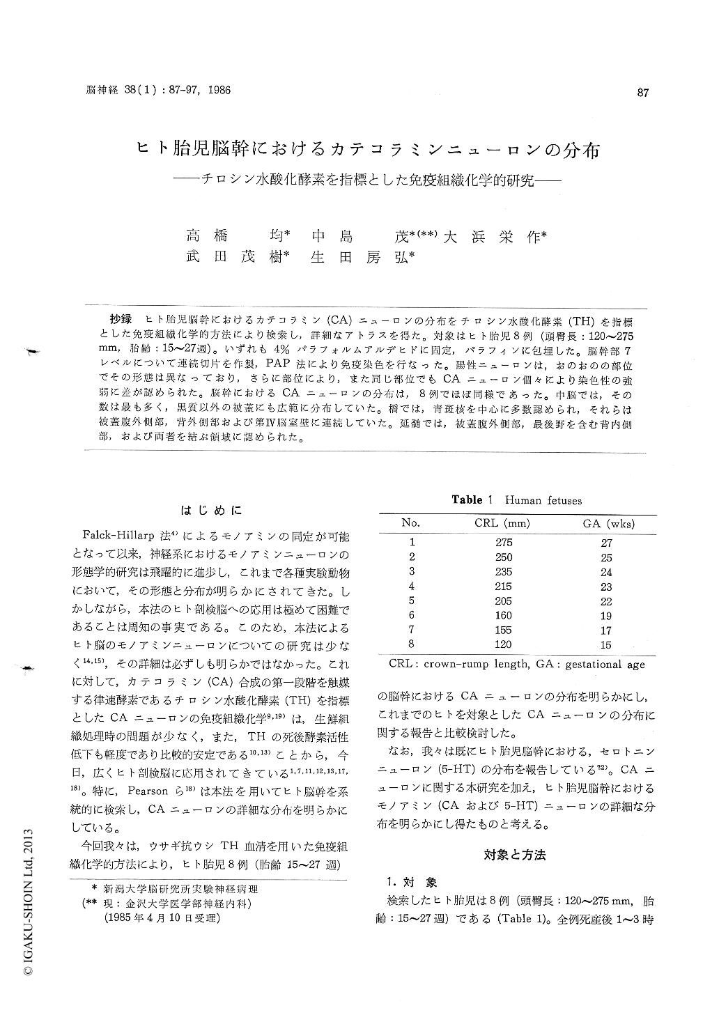
抄録 ヒト胎児脳幹におけるカテコラミン(CA)ニューロンの分布をチロシン水酸化酵素(TH)を指標とした免疫組織化学的方法により検索し,詳細なアトラスを得た。対象はヒト胎児8例(頭臀長:120〜275mm,胎齢:15〜27週)。いずれも4%パラフォルムアルデヒドに固定,パラフィンに包埋した。脳幹部7レベルについて連続切片を作製,PAP法により免疫染色を行なった。陽性ニューロンは,おのおのの部位でその形態は異なっており,さらに部位により,また同じ部位でもCAニューロン個々により染色性の強弱に差が認められた。脳幹におけるCAニューロンの分布は,8例でほぼ同様であった。中脳では,その数は最も多く,黒質以外の被蓋にも広範に分布していた。橋では,青斑核を中心に多数認められ,それらは被蓋腹外側部,背外側部および第IV脳室壁に連続していた。延髄では,被蓋腹外側部,最後野を含む背内側部,および両者を結ぶ領域に認められた。
Immunohistochemistry using antibodies to tyro-sine hydroxylase (TH), a rate-limiting enzyme which catalizes the initial step in the catechola-mine synthesizing pathway, has been widely ac-cepted as one of the methods for identification of catecholamine neurons in the nervous system.
In the present study, we performed immunohis-tochemical examination to elucidate the distribu-tion of catecholamine neurons in brain stem of human fetuses.
The brain stems were obtained from 8 human fetuses (CRL : 120~75 mm, GA : 15~27 wks) 1-3 h after death following therapeutic or sponta-neous abortion. They were immediately fixed with4% paraformaldehyde in 0.1 M phosphate buffer, pH 7.4, dehydrated with graded ethanol, and embedded in paraffin. Serial 6μm sections were cut from 7 different levels of the brain stem of each fetus. These sections were stained by pero-xidase-antiperoxidase (PAP) technique using TH antisera. The TH antisera used were raised in rabbits by injecting purified TH from bovine adrenal medulla. The preparation and the specifi-city of TH antisera were described in detail else-where (Nakashima et al, 1983).
Catecholamine neurons were clearly demonstra-ted in the brain stem of all fetuses. They could be recognized as catecholamine cell groups in the same manner as is done in experimental mammals. Among these cell groups, the catecholamine neu-rons showed distinct cytological features in shape and size. The distribution of catecholamine posi-tive neurons in the brain stem was almost the same in the 8 human fetuses, and an atlas was given with anatomical explanation under the ter-minology of Olszewski and Baxter (1982) for the human brain stem.
In the mesencephalon, a large number of cate-cholamine neurons lay in the nucleus substantiae nigrae, pars compacta, the nucleus paranigralis, the middle of the ventral tegmentum and the tractus tegmentalis centralis, and fewer catechola-mine neurons were scattered in the other tegmen-tal area. In addition, a group of small catechola-mine neurons was located in the griseum centrale mesencephali near the aqueduct. In the pons, catecholamine neurons occurred mainly in the nucleus locus coeruleus and the nucleus subcoeru-leus. A band of TH-posi tive neurons extended from the nucleus locus coeruleus to the dorsolate-ral tegmentum, and further to the roof of the fourth ventricle. Occasional catecholamine neurons were present in the area medial to the upper portion of the nucleus locus coeruleus. More caudally, a small number of catecholamine neurons were scattered in the area medial to the nervus facialis and adjacent to the nucleus facialis and the nucleus olivaris superior. In the medulla ob-longata, two groups of catecholamine neurons were recognized. These were located in the ventro-lateral and dorsomedial regions of the tegmentum. An oblique band of scattered catecholamine neurons bridged these two groups. The area postrema was revealed as a locus in which many small catecholamine neurons were located. With regard to the distribution of catecholamine neurons, our results were compared with the pre-vious studies made in the human CNS (Nobin and Bjorklund, 1973 ; Pearson et al 1983). The distribu-tion of catecholamine neurons demonstrated here was essentially similar to that of human adults de-scribed by Pearson et al (1983) although additional new observations were also pointed out.

Copyright © 1986, Igaku-Shoin Ltd. All rights reserved.


