Japanese
English
- 有料閲覧
PPV購入案内
1,320円 (1,200円+税10%) こちらは、117ページから126ページの文献です。購入には医書.jp本会員登録が必要です。
会員登録完了後に改めて上記「購入サイトへ」を選択してください。
または「購入サイトへ」選択後に「購入ログイン」、「新規会員登録」の順に進んでください。
会員登録について詳しくはをご確認ください。
- Abstract 文献概要
- 1ページ目 Look Inside
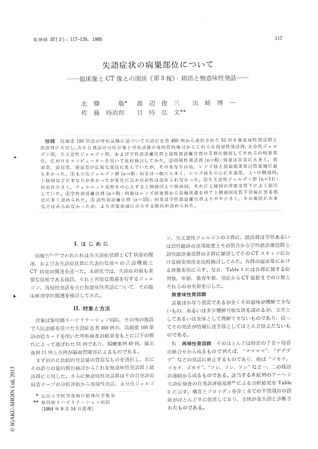
抄録 高頻度100単語の呼称試験に基づいて失語症患者450例から選択された51例を無意味性発話群と錯語群に大別し,次に自発語の分析評価と呼称試験の量的質的検討からこれらを再帰性発話群,未分化ジャルゴン群,失文意性ジャルゴン群,および字性錯語優位群と語性錯語優位群の5群に細別してそれらの病巣部位,広がりをコンピューターを用いて比較検討してみた。①再帰性発話群(n=8):病巣は非常に大きく,前頭葉,頭頂葉,側頭葉の広範な部位に及んでいたが,その重なりは島,レンズ核と前頭葉深部白質領域に最も多かった。②未分化ジャルゴン群(n=9):病巣は一般に大きく,レンズ核を中心に弁蓋部,上・中側頭回,上縁回などに重なりが多かったが変化に富み局在性は認められなかった。③失文意性ジャルゴン群(n=11):病巣は小さく,ウュルニッケ領野を中心とする上側頭回と中側頭回,それに上縁回の深部皮質下によく限局していた。④字性錯語優位群(n=8):病巣はレンズ核後部から島輪状溝を経て上側頭回皮質下領域に至る部位に多く認められた。⑤語性錆語優位群(n=15):病巣は字性錯語優位群よりやや小さく,その部位に大きな差はみられなかったが,より深部領域に分布する傾向が認められた。
According to the result of the visual naming task of 100 realistic pictures, 51 cases were selected from 450 cases with various types of aphasia and divided into two groups, namely, meaningless spe-ech group and paraphasic group. Subsequently, on the basis of the quantitative and qualitative valua-tion of the spontaneous speech and the naming task, the two groups were subdivided with mean-ingless speech group being recurring utterance (RU group), undifferentiated jargon (UJ group), aseman-tic jargon (AJ group) and paraphasic group being literal paraphasia (LP group), verbal paraphasia (VP group). Using a microcomputer, the locus and extent of the lesions, as demonstrated by computed tomography for each group were superimposed onto standardized matrices. The relationship between the focus and the extent of the lesions and the various groups was investigated.
1) RU group (n=8):The size of the lesions of this group was very large and almost all patients had extensive lesions involving frontal-temporal-parietal lobes. There was marked concentration ofthe lesions in the area of the insula, lenticular nucleus and the deep structures of the frontal lobe.
2) UJ group (n=9):In general, the size of the lesions was large and highly involved lesions were located in the lenticular nucleus and operculum as well as the superior and middle temporal gyri and the supramarginal gyrus. Because of the large variability in lesion patterns, it has proved to have little localizing value.
3) AJ group (n=11):The size of the lesions was significantly smaller than any other group. At least 80% of the patients had the superior temporal lesions involving Wernick's area and subcorticallesions of the supramarginal gyrus.
4) LP group (n=8) : The lesions were relatively large in size. While there was some concentration of the lesions in the area of the lenticular nucleus, posterior parts of the insula and anterior temporal gyrus, it was difficult to localize in a definite region.
5) VP group (n=15):The lesions of this group were a little smaller and located slightly deeper than those of the LP group. The finding has proved to have little localizing value.
Copyright © 1985, Igaku-Shoin Ltd. All rights reserved.