Japanese
English
- 有料閲覧
PPV購入案内
1,320円 (1,200円+税10%) こちらは、941ページから950ページの文献です。購入には医書.jp本会員登録が必要です。
会員登録完了後に改めて上記「購入サイトへ」を選択してください。
または「購入サイトへ」選択後に「購入ログイン」、「新規会員登録」の順に進んでください。
会員登録について詳しくはをご確認ください。
- Abstract 文献概要
- 1ページ目 Look Inside
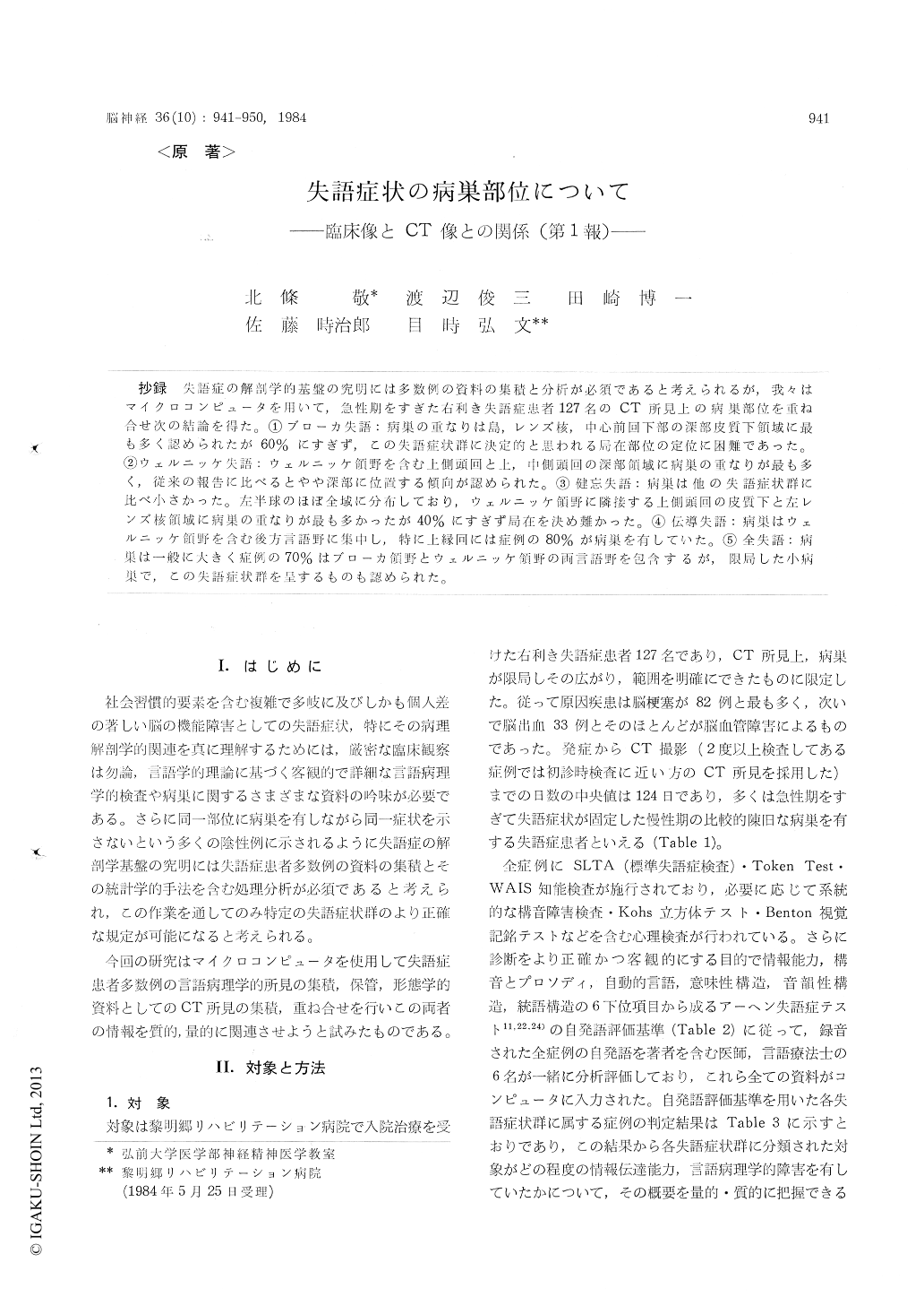
抄録 失語症の解剖学的基盤の究明には多数例の資料の集積と分析が必須であると考えられるが,我々はマイクロコンピュータを用いて,急性期をすぎた右利き失語症患者127名のCT所見上の病巣部位を重ね合せ次の結論を得た。①ブローカ失語:病巣の重なりは島,レンズ核,中心前回下部の深部皮質下領域に最も多く認められたが60%にすぎず,この失語症状群に決定的と思われる局在部位の定位に困難であった。②ウェルニッケ失語:ウェルニッケ領野を念む上側頭同と上,中側頭回の深部領域に病巣の重なりが最も多く,従来の報告に比べるとやや深部に位置する傾向が認められた。③健忘失語:病巣は他の失語症状群に比べ小さかった。左半球のほぼ企域に分布しており,ウェルニッケ領野に隣接する上側頭同の皮質下と左レンズ核領域に病巣の重なりが最も多かったが40%にすぎず局在を決め難かった。④伝導失語:病巣はウェルニッケ領野を含む後方言語野に集中し,特に上縁回には症例の80%が病巣を有していた。⑤全失語:病巣は一般に大きく症例の70%はブローカ領野とウェルニッケ領野の両言語野を包含するが,限局した小病巣で,この失語症状群を呈するものも認められた。
Using a microcomputer, the locus and extent of the lesions, as demonstrated by computed tomo-graphy for 127 cases with various types of aphasia were superimposed onto standardized marices. The relationship between the foci of the lesions and the types of aphasia was investigated.
1. Broca phasics (n=39):Since the accumulated site of the lesions highly involved the deep struc-tures of the lower part of the precentral gyrus as well as the insula and lenticular nucleus, only 60 % of the Broca aphasics had lesions on these areas. This finding has proved to have little loca-lizing value.
2. Wernicke aphasics (n=23):The size of the lesion was significantly smaller than Broca's apha-sia. At least 70% of the patients had the superior temporal lesions involving Wernicke's area and subcortical lesions of the superior and middle temporal gyri. The site of lesion corresponded roughly to the previous clinico-pathological reports but located a little deep.
3. Amnestic aphasics (n=18) : The size of the lesion was smaller than any other types. While there was some concentration of the lesions (ma-ximum 40%) in the area of the subcortical region of the anterior temporal gyrus adjacent to Wer-nicke's area and the lenticular nucleus, the lesions were distributed throughout the left hemisphere. Amnestic aphasia was thought to be the least localizable.
4. Conduction aphasics (n=11): The lesions were relatively small in size. Many patients had posterior speech area lesions involving at least partially Wernicke's area. In particular, more than 80% of the conduction aphasics had lesions of the supramarginal gyrus and it's adjacent deep struc-tures.
5. Global aphasics (n=36) : In general, the size of the lesion was very large and 70% of the glo-bal aphasics had extensive lesions involving both Broca's and Wernicke's areas. However, there were observations showing that the lesions can be small and confined.
Because of the large variability in lesion pat-terns and speech disturbances, it is necessary to expand the number of cases for relate detailed neuropsychological examinations with morpholo-gical CT-findings. Our method permits it easily to process and to analyze a large number of cases. By studying larger series, the better definition in the relationship between anatomic lesion location and aphasia type, even for less common aphasia syndromes could be obtained.
Copyright © 1984, Igaku-Shoin Ltd. All rights reserved.