Japanese
English
- 有料閲覧
PPV購入案内
1,320円 (1,200円+税10%) こちらは、413ページから417ページの文献です。購入には医書.jp本会員登録が必要です。
会員登録完了後に改めて上記「購入サイトへ」を選択してください。
または「購入サイトへ」選択後に「購入ログイン」、「新規会員登録」の順に進んでください。
会員登録について詳しくはをご確認ください。
- Abstract 文献概要
- 1ページ目 Look Inside
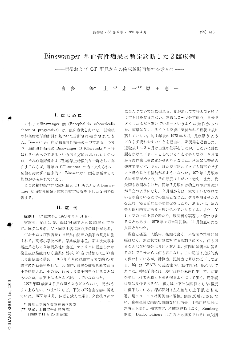
I.はじめに
これまでBinswanger病(Encephalitis subcorticalischronica progressiva)は,臨床症状とあわせ,剖検後の神経病理学的所見に基づいて診断され報告されてきた。Binswanger病が脳血管性痴呆の一型である,つまり,脳血管性痴呆のBinswanger型(Olszewski)6)と呼ばれるべきものであるという考え方にわれわれは立つが,それが臨床像および形態学上特徴的な一群として存在するならば,近年のCT scannerの力に支えられて,剖検を待たずに臨床的にBinswanger型を診断する可能性がひらかれていよう。
ここに精神医学的な臨床像とCT所見とからBinswa—nger型血管性痴呆と臨床的暫定診断を下した2例を報告する。
The diagnosis of subcortical arteriosclerotic en-cephalopathy of Binswanger should be comfirmed neuropathologically by postmortem examination. But we expect that it may be possible to diagnose provisionally as Binswanger's disease on the basis of clinical course and findings including computed tomography.
Two cases, 57 years old postman (case 1) and 60 years old housewife (case 2) were reported. They had hypertension for over 20 years in their past history and showed a progressive dementia since before one year and 9 years. Transient syncopal attack and epileptiform seizure revealed occasional-ly. Psychiatrically either of them were apathetic, aspontaneous and autistic. One of them (case 2) showed a striking paranoid-hallucinatory state. Spas-tic gait disturbance and dysarthria are found neu-rologically, and moreover in case 1 revealed the incontinence of urine and feces. Arteriosclerotic changes were seen in the fundi. The EEG showed a slow α rhythm with scattered θ- and δ-waves. Labolatory data of blood, urine and CSF were normal.
On the basis of these clinical course and findings the diagnosis of Binswanger's type of cerebral ar-teriosclerosis were suspected. The CT-scan of 2 cases proved a symmetrical enlargement of the lat-eral ventricles and marginated areas of definite abnormal low density in the white matter of the occipital (case 1) and frontal (case 1 and 2) lobes. Conclusively we may provisionally diagnosed our two cases as Binswanger's disease, also supported by the findings of CT.
Copyright © 1981, Igaku-Shoin Ltd. All rights reserved.