Japanese
English
- 有料閲覧
PPV購入案内
1,320円 (1,200円+税10%) こちらは、985ページから993ページの文献です。購入には医書.jp本会員登録が必要です。
会員登録完了後に改めて上記「購入サイトへ」を選択してください。
または「購入サイトへ」選択後に「購入ログイン」、「新規会員登録」の順に進んでください。
会員登録について詳しくはをご確認ください。
- Abstract 文献概要
- 1ページ目 Look Inside
I.はじめに
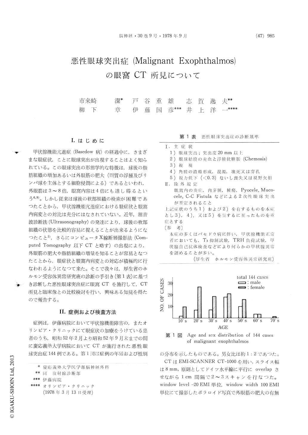
甲状腺機能亢進症(Basedow 病)の経過中に,さまざまな眼症状,ことに眼球突出が出現することはよく知られている。この眼球突出の形態学的な特徴は,球後の脂肪組織の増加あるいは外眼筋の肥大(間質の浮腫及びリンパ球を主体とする細胞侵潤による)であるといわれ,外眼筋は3〜8倍,眼窩内容は4倍にも達し得るという6,9)。しかし従来は球後の軟部組織の検索が困難であつたことから,甲状腺機能亢進症における眼症状と眼窩内病変との対比は充分にはなされていない。近年,超音波診断法(Ultrasonography)の発達により,球後の軟部組織の状態を比較的容易に捉えることが出来るようになつたこと2),さらにコンピュータX線断層撮影法(Com—puted Tomography以下CTと略す)の出現により,外眼筋の肥大や脂肪組織の増量を知ることが容易となつたことから,眼症状と眼窩内病変との対応が積極的に行なわれるようになつて来た。そこで我々は,厚生省のホルモン受容体異常研究班の診断の手引き(第1表)に基づき診断した悪性眼球突出症に眼窩CTを施行して,CT所見と臨床像との比較検討を行い,興味ある知見を得たので報告する。
The following is a report on the comparative study of the ophthalmological symptoms and the orbital CT scan findings of malignant exophthalmos that were diagnosed by the established criteria of the Ministry of Health and Welfare. A total of 144 malignant exophthalmos that underwent CT scan at the Keio University Hospital in the 8 months from February to September 1977 were studied.
For orbital CT scan EMI-SCANNER CT-1000 was employed at a slice width of 8mm to take as a rule 2-3 scans at 1 cm interval with a parallel overlap on the Reid's baseline. The presence of the enlargement of the extraocular muscle wasdetermined on polaroid photographs obtained at window level of -20 EMI units and window width of 100 EMI units.
The Orbital CT scan findings of malignant ex-ophthalmos can be classified into those that reveal characteristic enlargement of the extraocular muscle and those that suggest increase in the retrobulbar fat tissue. The enlargement of the extraocular muscle was detected in 87 (60.4%) out of the 144 cases. The remaining 57 cases were considered to be due to the increase in the retrobulbar fat tissue. Hence, when possible causative lesions for exoph-thalmos are not detected on CT scan of patients with exophthalmos the presence of malignant ex-ophthalmos should be suspected.
Of the extraocular muscles the enlargement of the inferior rectus muscle was the most frequent (46.5%) followed by the medial rectus muscle (29.9 %) and the lateral rectus muscle (20.1%). Accord-ingly, the CT scan of malignant exophthalmos should be so devised to expose the inferior portion of the orbit in order to determine the presence of the inferior rectus muscle.
The muscle which caused the disturbance in the eye ocular movement often showed enlargement on CT scan study. The degree of the enlargement and the extent of the disturbance of the ocular movement, however, did not necessarily correspond with each other. Cases with visual disturbance often showed findings suggestive of optic nerve compression by the extraocular muscle enlarged at the apex of the orbit.
Unilateral malignant exophthalmos was detected in only 7 cases (4.9%). Even in the unilateral cases the changes in the extraocular muscle were often bilateral under CT scan.
Copyright © 1978, Igaku-Shoin Ltd. All rights reserved.