Japanese
English
- 有料閲覧
- Abstract 文献概要
- 1ページ目 Look Inside
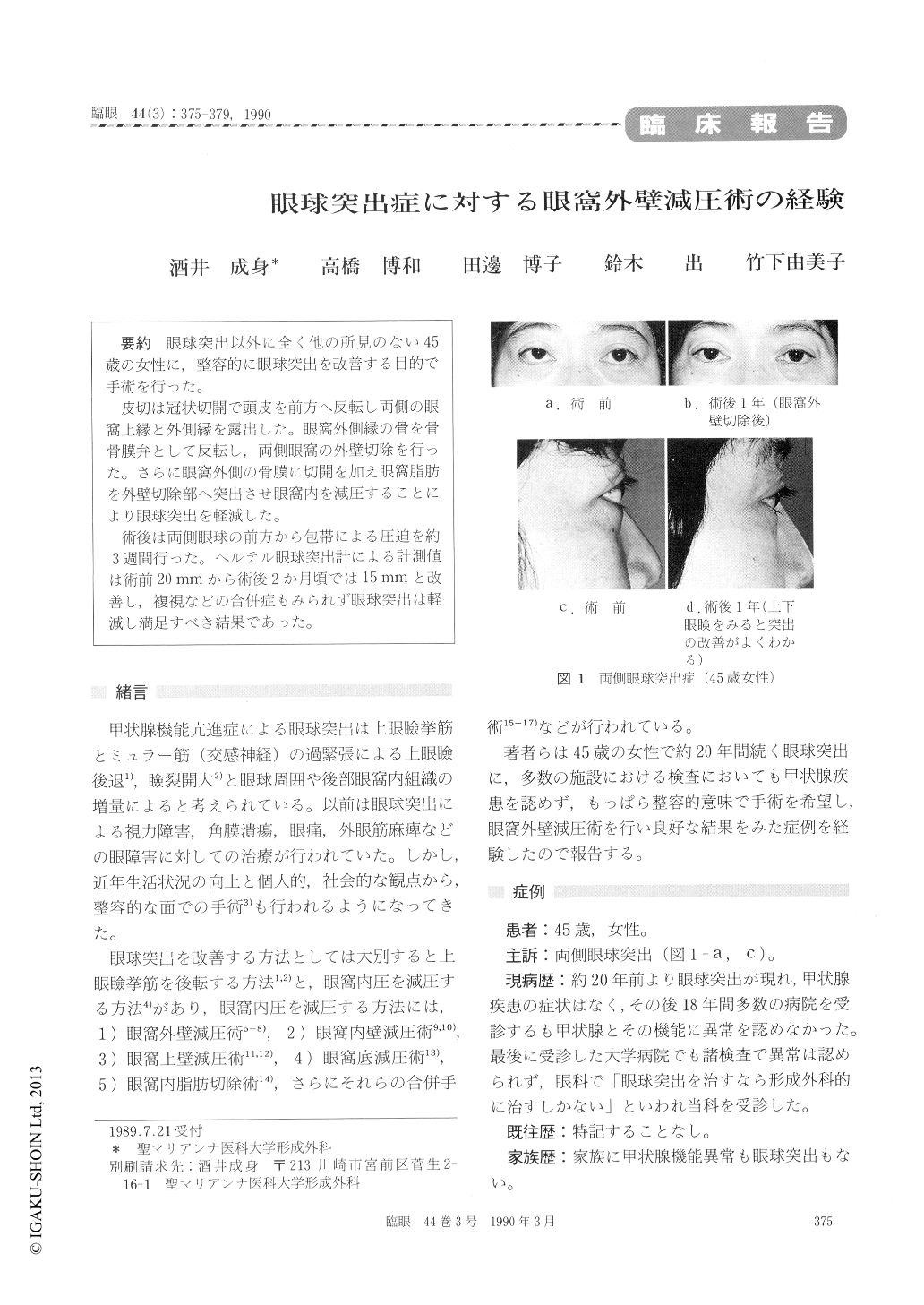
眼球突出以外に全く他の所見のない45歳の女性に,整容的に眼球突出を改善する目的で手術を行った。
皮切は冠状切開で頭皮を前方へ反転し両側の眼窩上縁と外側縁を露出した。眼窩外側縁の骨を骨骨膜弁として反転し,両側眼窩の外壁切除を行った。さらに眼窩外側の骨膜に切開を加え眼窩脂肪を外壁切除部へ突出させ眼窩内を減圧することにより眼球突出を軽減した。
術後は両側眼球の前方から包帯による圧迫を約3週間行った。ヘルテル眼球突出計による計測値は術前20mmから術後2か月頃では15mmと改善し,複視などの合併症もみられず眼球突出は軽減し満足すべき結果であった。
A 45-year-old female presented with bilateral exophthalmos of 20 years' duration. Thyroid dysfunction was ruled out through systemic and laboratory examinations. The exophthalmos mea- sured 20mm hertel for either eye. We performed decompression surgery of the orbit through lateral orbital approach. Following coronary incision on the forehead, the forehead skin flap was turned down to midface until exposure of the upper andlateral orbital rim. After removal of the lateral osseous orbital wall, we severed the orbital periost to induce protrusion of orbital fat tissue responsible for the exophthalmos. After sufficient amount of fat tissue protruded, the surgical wound was closed in the reverse order.
The postsurgical course was uneventful and satis-factory. The amount of proptosis decreased to 15 mm hertel 2 months after surgery. This procedure is recommended for its safety and effectiveness.

Copyright © 1990, Igaku-Shoin Ltd. All rights reserved.


