Japanese
English
- 有料閲覧
- Abstract 文献概要
- 1ページ目 Look Inside
I.はじめに
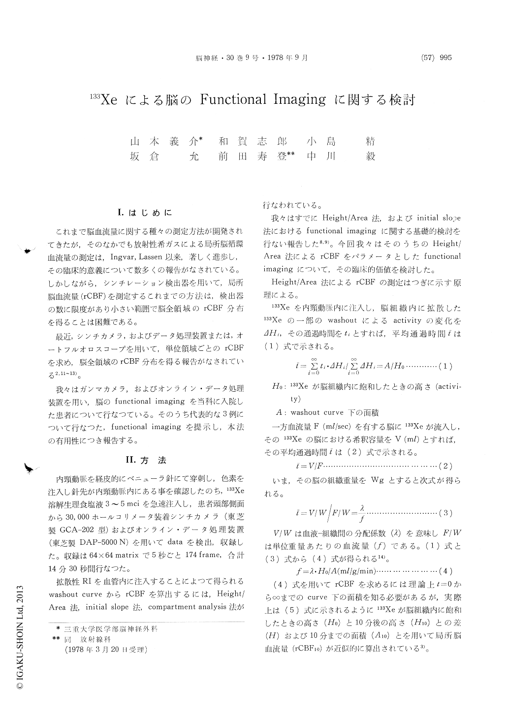
これまで脳血流量に関する種々の測定方法が開発されてきたが,そのなかでも放射性希ガスによる局所脳循環血流量の測定は,Ingvar,Lassen以来,著しく進歩し,その臨床的意義について数多くの報告がなされている。しかしながら,シンチレーション検出器を用いて,局所脳血流量(rCBF)を測定するこれまでの方法は,検出器の数に限度があり小さい範囲で脳全領域のrCBF分布を得ることは困難である。
最近,シンチカメラ,およびデータ処理装置または,オートフルオロスコープを用いて,単位領域ごとのrCBFを求め,脳全領域のrCBF分布を得る報告がなされている2,11〜13)。
Since Ingver and Lassen measured regional cere-bral blood flow (rCBF) first in 1963, measurement of rCBF and its clinical application have widely been performed in the world.
In this paper the authors show practical value of the brain functional imaging of rCBF10 measured by Height/Area method using 133Xe.
The joint camera-computer system used consists of the GCA-202 scinticamera and the DAP-5000N computer system. Following internal carotid in-jection of 3-5 mCi of 133Xe, 5second frames of data on a 64×64 matrix are obtained for a period of 14.5 minutes, utilizing the on-line computer system. The method is able to make a functional image of rCBF10 by means of subtracting some amounts of rCBF10 from the original rCBF10 and the affected areas become more clear in photographs. For ex-ample, when the maximum rCBF10 is 41 ml/100gr/ min and the average rCBF10 is 21 ml/100gr/min, the areas where rCBF10 is below 10ml/100gr/min are neglected and showed on the photographs. This method can clearly show the areas with decreased rCBF and is routinely used in our department in cases with cerebral vascular disease.
In this paper the authors represent typical cases with cerebral infarction, Neuro-Behcet and arterio-venous malformation (AVM).
Functional image of rCBF10 in a case with cere-bral infarction clearly showed the areas with de-creased rCBF10 and that of a case with AVM demon-strated that rCBF10 was normalized near the region of the AVM after total removal where rCBF had been abnormally high. Functional image in a case with Neuro-Behçet syndrome immediately after ischemic insult revealed remarkably increased areas of rCBF10 where cerebral angiography also showed capillary blush and early filling veins.
Functional imaging of rCBF is very reliable method and clear at a glance for detecting the lesion and effects of treatment in cerebral vascular disease.

Copyright © 1978, Igaku-Shoin Ltd. All rights reserved.


