Japanese
English
症例報告
頭蓋底に見られた海綿状血管腫の1剖検例
CAVERNOMA ON THE INTRACRANIAL BASE: REPORT OF A CASE
小笠原 定則
1
,
中沢 一臣
1
,
桂 佐元
1
,
大泉 早苗
2
Sadanori Ogasawara
1
,
Kazuomi Nakazawa
1
,
Sukemoto Katsura
1
,
Sanae Oizumi
2
1岩手医科大学医学部病理学第2講座
2岩手労災病院内科
1Department of Pathology II, School of Medicine, Iwata Medical University
2Iwate Rosai Hospital
pp.735-737
発行日 1973年6月1日
Published Date 1973/6/1
DOI https://doi.org/10.11477/mf.1406203335
- 有料閲覧
PPV購入案内
1,320円 (1,200円+税10%) こちらは、735ページから737ページの文献です。
PPV(ペイ・パー・ビュー)とは、論文(記事)単位で購入・閲覧ができるサービスです。
購入には医書.jp本会員登録が必要です。
会員登録完了後に改めて上記「購入サイトへ」を選択してください。
または「購入サイトへ」選択後に「購入ログイン」、「新規会員登録」の順に進んでください。
会員登録について詳しくはをご確認ください。
購入には医書.jp本会員登録が必要です。
会員登録完了後に改めて上記「購入サイトへ」を選択してください。
または「購入サイトへ」選択後に「購入ログイン」、「新規会員登録」の順に進んでください。
会員登録について詳しくはをご確認ください。
- Abstract 文献概要
- 1ページ目 Look Inside
はじめに
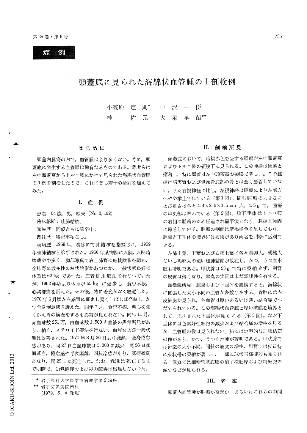
頭蓋内腫瘍の内で,血管腫は余り多くない。特に,頭蓋底に発生する血管腫は稀有なるものである。著者らは左中頭蓋窩からトルコ鞍にかけて見られた海綿状血管腫の1例を剖検したので,これに関し若干の検討を加えてみた。
The case was a 64-year-old miner, who died of silicotuberculosis. He had experienced neither cerebral nor endocrine disturbances. Unexpectedly, autopsy disclosed an encapsulated tumor extending from the sella turcica to the left middle cranial fossa. The tumor was completely covered with the dura mater, and adhered firmly to it. The hypophysis is compressed to the right with the tumor, it was, however, clearly separated from the hypophysis with the fibrous tissue. The tumor weighed 4.5g, and 4.5 × 2.5 × 1.5cm in size.
Copyright © 1973, Igaku-Shoin Ltd. All rights reserved.