Japanese
English
- 有料閲覧
- Abstract 文献概要
- 1ページ目 Look Inside
緒言
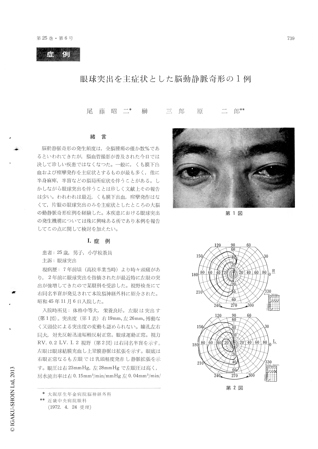
脳動静脈奇形の発生頻度は,全脳腫瘍の僅か数%であるといわれてきたが,脳血管撮影が普及された今日では決して珍しい疾患ではなくなつた。一般に,くも膜下出血および痙攣発作を主症状とするものが最も多く,他に半身麻痺,半盲などの脳局所症状を伴うことがある。しかしながら眼球突出を伴うことは珍しく文献上その報告は少い。われわれは最近,くも膜下出血,痙攣発作はなくて,片眼の眼球突出のみを主症状としたところの大脳の動静脈奇形症例を経験した。本疾患における眼球突出の発生機構については殊に興味ある所であり本例を報告してこの点に関して検討を加えたい。
A case of exophthalmos resulting from supara-tentorial arteriovenous malformation, is reported. The patient was a 29 year-old male with a com-plaint of slight headache of several years standing.Two years ago, his mother noticed a protrusion of his left eye, and this had gradually become more pronounced recently. On Nov. 6, 1970, he visited our hospital.
Ophthalmic examinations revealed unilateral exo-phthalmos with values of 26 mm for the left eye as compared to 19 mm for the right eye on the Hertel Exophthalmometer, and right homonymous hemianopsia and high intraocular pressure of the left eye, which was 28 mmHg on the Applanation Tonometer. The left ocular fundus showed a normal disc but dilatation of retinal veins. No other neuro-logic findings were observed. Serial carotid angio-graphy showed a large arteriovenous malformationin left post-temporal region. Radio-opaque material was observed in the cavernous sinus 2 seconds after injection of 60% Conray into the common carotid artery and was still visible after 6 seconds. This suggests congestion of the cavernous sinus and superior ophthalmic vein which may be cause of the protrusion of the left eye.
On December 4, 1970, surgical ligation of the left common carotid artery was performed. Post-operatively, exophthalmos of the left eye did not diminish, but intraocular pressure decreased and has remained within normal range for one year.
Copyright © 1973, Igaku-Shoin Ltd. All rights reserved.