Japanese
English
- 有料閲覧
PPV購入案内
1,320円 (1,200円+税10%) こちらは、953ページから957ページの文献です。購入には医書.jp本会員登録が必要です。
会員登録完了後に改めて上記「購入サイトへ」を選択してください。
または「購入サイトへ」選択後に「購入ログイン」、「新規会員登録」の順に進んでください。
会員登録について詳しくはをご確認ください。
- Abstract 文献概要
- 1ページ目 Look Inside
緒言
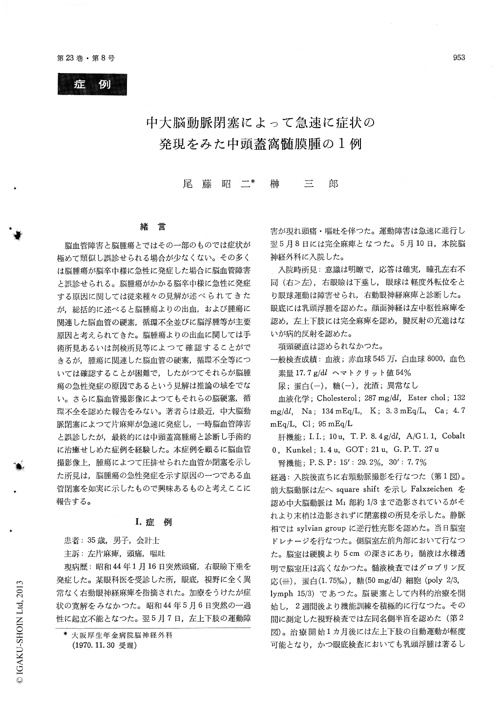
脳血管障害と脳腫瘍とではその一部のものでは症状が極めて類似し誤診せられる場合が少なくない。その多くは脳腫瘍が脳卒中様に急性に発症した場合に脳血管障害と誤診せられる。脳腫瘍がかかる脳卒中様に急性に発症する原因に関しては従来種々の見解が述べられてきたが,総括的に述べると脳腫瘍よりの出血,および腫瘍に関連した脳血管の硬塞,循環不全並びに脳浮腫等が主要原因と考えられてきた。脳腫瘍よりの出血に関しては手術所見あるいは剖検所見等によつて確認することができるが,腫瘍に関連した脳血管の硬塞,循環不全等については確認することが困難で,したがつてそれらが脳腫瘍の急性発症の原因であるという見解は推論の域をでない。さらに脳血管撮影像によつてもそれらの脳硬塞,循環不全を認めた報告をみない。著者らは最近,中大脳動脈閉塞によつて片麻痺が急速に発症し,一時脳血管障害と誤診したが,最終的には中頭蓋窩腫瘍と診断し手術的に治癒せしめた症例を経験した。本症例を顧るに脳血管撮影像上,腫瘍によつて圧排せられた血管が閉塞を示した所見は,脳腫瘍の急性発症を示す原因の一つである血管閉塞を如実に示したもので興味あるものと考えここに報告する。
A thirty-five year old man who had a right blepharoptosis for four months, was admitted to our clinic because of the sudden onset of a left hemiparesis with complaints of headache and Tomit-ting, but his consciousness was not impaired. The neurologic examination showed a paresis of the right oculomotor nerve, left facial nerve and left ex-tremities. The right carotid angiography, performed immediately after admission, showed occlusion of right middle cerebral artery in the middle of the M-I section. We diagnosed this case as a cerebral infarction and treated him. One month later, the 2nd right carotid angiography showed the complete disapperance of the occlusion and upward displace-ment and streching of the M-I section of the right middle cerebral artery. The brain scintigram revealed a concentration of 131I in the right tem-poral region. It was diagnosed as a right middle fossa tumor and suspicion of meningioma. When operated on, the tumor was located in the right middle fossa and directly under the M-I section of the middle cerebral artery and extended to the parasellar region, and was totally extirpated. The histological findings of the tumor showed menin-gioma.
Copyright © 1971, Igaku-Shoin Ltd. All rights reserved.