Japanese
English
- 有料閲覧
- Abstract 文献概要
- 1ページ目 Look Inside
はじめに
慢性硬膜下血腫は脳神経外科領域において最も普遍的な疾患の1つであり,研究の歴史も古い14)15)。
慢性硬膜下血腫に関する従来の報告を通覧し,今日なお残されている問題を集約すると次の3項目を挙げることができよう。
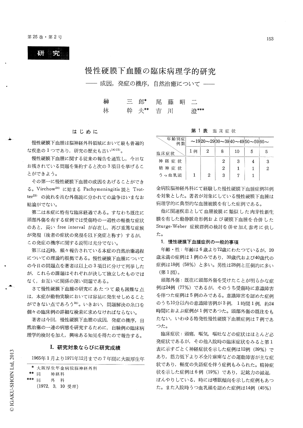
We studied clinicopathological examination onthe 31 cases with chronic subdural hematoma and 2 cases with analogous disease who were admitted to our clinic from 1965 to 1971. All cases were treated surgically. Twenty-nine of 31 cases with chronic subdural hematoma were treated by flap craniotomy and 2 were by burr hore. Twenty-four cases had a history of trivial head trauma but 6 had no trauma.
We examined the clinical course of each cases in detail i. e. a history of head trauma, a term of free interval, onset or recurrence of symptoms, data of lumbar puncture, findings of ocular fundus and neuroradiologic examination, and then we com-pared them with pathological findings of hematoma membranes taken at operation.
The results obtained in this study were as follows,
1) The histopathological age of hematoma mem-brane in each case was more corresponding to the period from the manifestation of symptoms to opera-tion than to the period from initial head trauma to operation. From these findings we considered that the development of hematoma membrane should not start at the time immediately after the head trauma. Subdural hemorrhage from ruptured bridg-ing veins following trauma would be easily ab-sorbed in almost all cases but subdural hemorrhage might stimulate the dura mater to react excessively in a few cases which must be induced by certain factors such as alcoholism, age and vitamine C deficiency regarding these abnormal conditions. The reaction of dura mater seemed to produce a new granulation membrane, which was demon-strated in our own cases with ruptured arterio-venous malformation. This new membrane consisted of fibrous tissue and abundant macrocapillary called as sinusoidal channel in chronic subdural hematoma. In our opinion subdural new membrane proliferate insidiously during two or three months or longer, and if intramural hemorrhage occures the new membrane will proceed into hematoma.
2) The pressure of cerebrospinal fluid of each case had no intimate relation with the amount of hematoma. The pressure of cerebrospinal fluid decreased rapidly after evacuation of hematoma in the cases with not so high intracranial pressure without papilla edema. While the pressure de-creased gradually for two or three weeks after the evacuation of hematoma in the cases with remark-able high intracranial pressure with papilla edema. The difference in decreasing the intracranial pres-sure would be due to brain edema. Brain edema is probably pathophysiologic basis for abnormal clinical features such as papilla edema, hemiplegia etc.
3) It was demonstrated pathologically in this study that the hematoma membrane might proceed from proliferating stage to degenerating stage in its life history. From the view point of histological findings of the hematoma membrane it would be reasonable that chronic subdural membrane might be resorbed without operative procedure, provided that the brain edema beneath the hematoma which manifested clinical features were controlled by osmo-therapy.
Copyright © 1973, Igaku-Shoin Ltd. All rights reserved.