Japanese
English
- 有料閲覧
PPV購入案内
1,320円 (1,200円+税10%) こちらは、375ページから380ページの文献です。購入には医書.jp本会員登録が必要です。
会員登録完了後に改めて上記「購入サイトへ」を選択してください。
または「購入サイトへ」選択後に「購入ログイン」、「新規会員登録」の順に進んでください。
会員登録について詳しくはをご確認ください。
- Abstract 文献概要
- 1ページ目 Look Inside
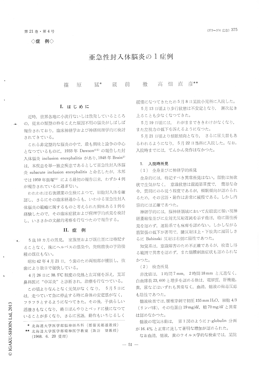
I.はじめに
近時,世界各地に小流行ないしは散発しているところの,従来の類型の枠をこえた原因不明の脳炎がしばしば報告されており,臨床神経学および神経病理学的に検討されてきている。
これら非定型的な脳炎の中で,最も興味と論争の中心となつているものに,1933年Dawson4)5)の報告した封入体脳炎inclusion encephalitisがあり,1948年Brain2)は,本疾患を単一独立疾患であるとして亜急性封入体脳炎subacute inclusion encephalitisと命名したが,本邦では1959年飯塚11)による最初の報告以来,わずか4例が報告されているに過ぎない。
1) This is a 5-year-old boy of subacute ence-phalitis (Dawson's inclusion encephalitis) confirmed by right occipital lobe biopsy.
2) The onset of illness was noted by loss of left visual acuity and impaired consciousness.
The neurological examination on admission revealed impaired consciousness (confusion grade II), loss of left visual acuity, absence of left light reflex, left extensor plantar reflex and hypotonia of all extre-mities.
The clinical course was characterized by progressive impairment of consciousness, ballismus-like move-ment of upper extremities, and alternate appearance of decorticate or decerebrate posture.
2) The cerebrospinal fluid was normal except for increased γ-glohuline on electrophoresis.
The complement fixation test and isolation of viru-ses from the cerebrospinal fluid and feces were negative.
4) The right occipital lobe biopsied on the 25th hospital day revealed as follow :
Macroscopically, the meninges and cerebral cortex were normal.
Microscopically, perivascular cellular infiltration, de-generation of nerve cells, proliferation of microglia and intranuclear inclusion bodies of Cowdry's A-type were demonstrated.
There was no hemorrhage or necrotic lesion.
5) This case was discussed comparing with other references.
Copyright © 1969, Igaku-Shoin Ltd. All rights reserved.