Japanese
English
- 有料閲覧
- Abstract 文献概要
- 1ページ目 Look Inside
I.はじめに
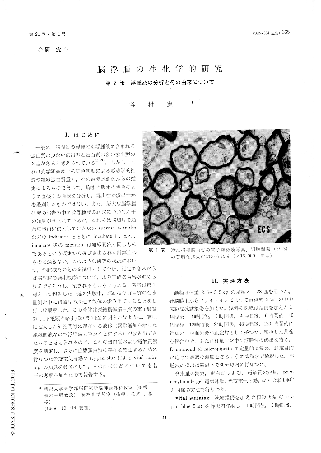
一般に,脳間質の浮腫にも浮腫液に含まれる蛋白質の少ない漏出型と蛋白質の多い滲出型の2型があると考えられている1)〜3)。しかし,これは光学顕微鏡上の染色態度による形態学的推論や組織蛋白質量や,その電気泳動像からの推定によるものであつて,胸水や腹水の場合のように直接その性状を分析し,漏出性か滲出性かを鑑別したものではない。また,膨大な脳浮腫研究の報告の中には浮腫液の組成について若干の知見が含まれているが,これらは脳切片を通常細胞内に侵入していかないsucroseやinulinなどのindicatorとともにincubateし,かつ,incubate後のmediumは組織醐液と同じものであるという仮定から導びき出された計算上のものに過ぎない。このような研究の現況において,浮腫液そのものを試料として分析,測定できるならば脳浮腫の発生機序について,より正確な考察が進められるであろうし,望まれるところでもある。著者は第1報として報告した一連の実験中,凍結損傷群白質の含水量測定中に組織片の川辺に液体の滲み出てくることをしばしば観察した。この液体は凍結損傷脳白質の電子顕微鏡(以下電顕と略す)像(第1図)に明らかなように,著明に拡大した細胞間隙に存在する液体(異常増加を示した組織間液なので浮腫液と呼ぶことにする)が滲み出てきたものと考えられるので,これの蛋白質および電解質濃度を測定し,さらに血漿蛋白質の存在を確認するために行なつた免疫電気泳動やtrypan blueによるvltal stain—ingの知見を参考にして,その由来などについても若干の考察を加えたので報告する。
In this series, an appearance of fluid of the edema-tous white matter dissected from the cold injured brain was observed. Marked enlargement of ex-tracellular space filled with osmophilic substance and the swelling of astrocytic processes were revealed by electronmicroscopic investigation. This fluid was responsible for abnormal increase in interstitial fluid of brain tissue.
Experimental brain edema was produced in adult cats using the freezing technique. Fresh edematous white matter was collected in tightly sealed small phial, then edema fluid appeared spontaneously was isolated with Drummond's micropipette. These pro-cedures were carried out at room temeprature within 30 minutes after the sacrifice. This resulted in the separation of a fluid of about 100μl from edematous white matter of 300-500 mg. Biochemical investiga-tion, i. e., determination of electrolytes and protein content, polyacrylamide gel electrophoretic and im-munoelectrophoretic analyses were carried out with this material by the same technique described in the previous publication, and compared with those of serum. In order to get a reasonable interpretation of the origin of edema fluid, trypan blue in a dose of 5 ml of 5 per cent solution was intravenously injected shortly after the application of solid CO2.
The results were : (1) When water content in-creased by 5 per cent more than control value, it was possible to obtain edema fluid. Actually, at 10, 12, 24, 48 hours after production of the lesion, edema fluid could be collected. (2) Compared with serum, edema fluid contained significantly less sodium, quite high potassium and equal chloride in their concentra-tions. Because the concentration of (Na+K) in the edema fluid was equal to that of serum, it could be considered that the fluid was osmotically isotonic to serum with respect to these electrolytes. High con-centration in potassium and low concentration in sodium in the edema fluid may be due partially to the dysfunction of cation transport system of mem-brane in vivo and to the preparation procedure em-ployed. (3) Protein concentration of the fluid re-ached the serum protein level within 24 hours after production of the lesion. Electrophoretic pattern of the edema fluid was similar to that of serum, though strictly speaking, there were some differences in that several bands migrated more rapidly than albu-min. With respect to this point, the edema fluid accords with the soluble protein extracted from ede-matous white matter of the cold injury group. Namely, the edema fluid was a mixture of serum and soluble cerebral protein. It has been responsible for a move of soluble protein from the affected cor-tex. This interpretation may be supported by the present experiment in that edematous white matter was gradually stained in trypan blue begining from adjacent area of deeply stained cortical lesion to fur-ther area of experimental hemisphere. Specific bind-ing of this dye to albumin was believed to be due to electrostatic interaction between the dye and ly-sine residues in the protein and soluble cerebral protein might move as stained albumin in the ede-matous brain tissue. (4) Immunoelectrophoretic analysis revealed the constituent of plasma proteins entered into the edematous brain. Usually precipita-tion lines dected in the edema fluid were fewer than in the serum obtained from the same animal, es-pecially in the region of alpha-2, and beta-1. This tendency was more distinct in the early stage of brain edema.
Copyright © 1969, Igaku-Shoin Ltd. All rights reserved.