Japanese
English
- 有料閲覧
PPV購入案内
1,320円 (1,200円+税10%) こちらは、1167ページから1171ページの文献です。購入には医書.jp本会員登録が必要です。
会員登録完了後に改めて上記「購入サイトへ」を選択してください。
または「購入サイトへ」選択後に「購入ログイン」、「新規会員登録」の順に進んでください。
会員登録について詳しくはをご確認ください。
- Abstract 文献概要
- 1ページ目 Look Inside
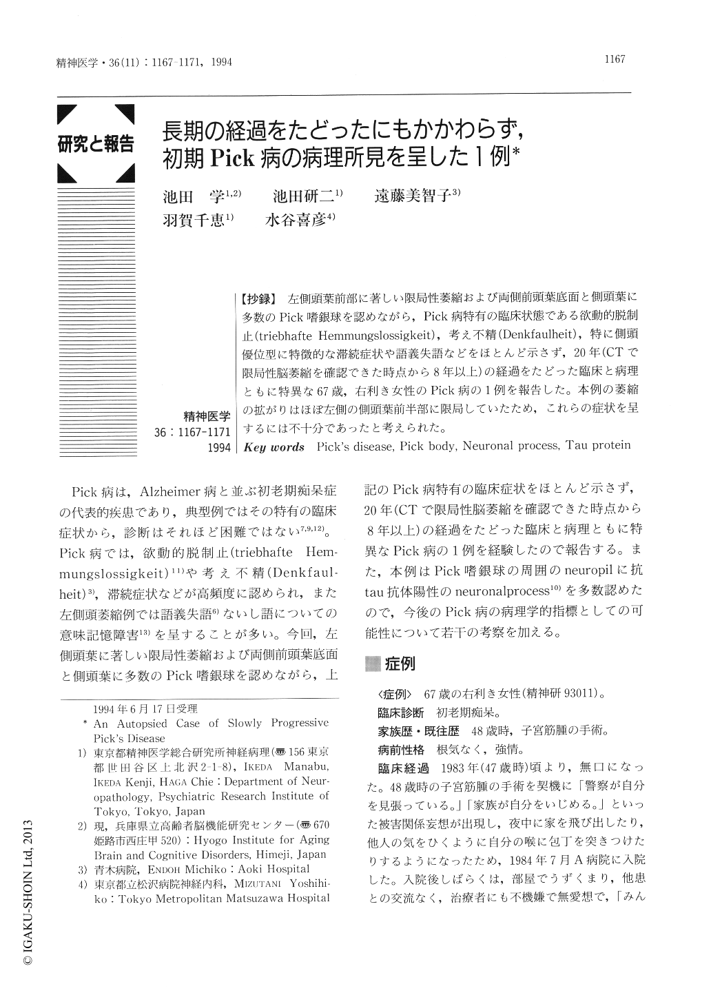
【抄録】 左側頭葉前部に著しい限局性萎縮および両側前頭葉底面と側頭葉に多数のPick嗜銀球を認めながら,Pick病特有の臨床状態である欲動的脱制止(triebhafte Hemmungslossigkeit),考え不精(Denkfaulheit),特に側頭優位型に特徴的な滞続症状や語義失語などをほとんど示さず,20年(CTで限局性脳萎縮を確認できた時点から8年以上)の経過をたどった臨床と病理ともに特異な67歳,右利き女性のPick病の1例を報告した。本例の萎縮の拡がりはほぼ左側の側頭葉前半部に限局していたため,これらの症状を呈するには不十分であったと考えられた。
A 67-year-old right-handed woman had a history of slowly progressive dementia. At the age of 48 she transiently experienced delusions, and was admitted to a mental hospital. During the subsequent 19 years, her predominant symptoms were personality changes such as euphoria and puerilism. At about 62 year of age, she started to lose spontaneity and develop a mild anomia. CT scan at age 59 revealed bilateral anterior temporal atrophy which was accentuated on the left side (Fig. 1). During her clinical course, “triebhafte Hemmungslossigkeit”, “stehende Symptome”, and “Gogi (word meaning) aphasia”, which are the characteristic symptoms of temporal dominant type of Pick's disease, were not detected.
Neuropathologically, atrophy was mostly circumscribed in the left anterior temporal lobe (Fig 2). Microscopically, mild nerve cell loss with tissue rarefacation and subcortical gliosis were found in the anterior temporal lobes, especially on the left side, and, in lesser degree, also in the anterior parts of the orbital, rectal, cingulate, and insular gyri (Fig. 3, 4). Silver impregnations revealed numerous Pick's argentophilic bodies (PBs) in the abovementioned regions. All the PBs were intensely stained with anti-tau antibody (Fig. 5a). In addition, many tau-positive granules (neuronal processes named by Murayama) were visualized in the neuropil around PB -bearing cells (Fig. 5b). These granules were hard to detect by routine silver stainings.
Copyright © 1994, Igaku-Shoin Ltd. All rights reserved.