Japanese
English
- 有料閲覧
PPV購入案内
1,320円 (1,200円+税10%) こちらは、559ページから563ページの文献です。購入には医書.jp本会員登録が必要です。
会員登録完了後に改めて上記「購入サイトへ」を選択してください。
または「購入サイトへ」選択後に「購入ログイン」、「新規会員登録」の順に進んでください。
会員登録について詳しくはをご確認ください。
- Abstract 文献概要
- 1ページ目 Look Inside
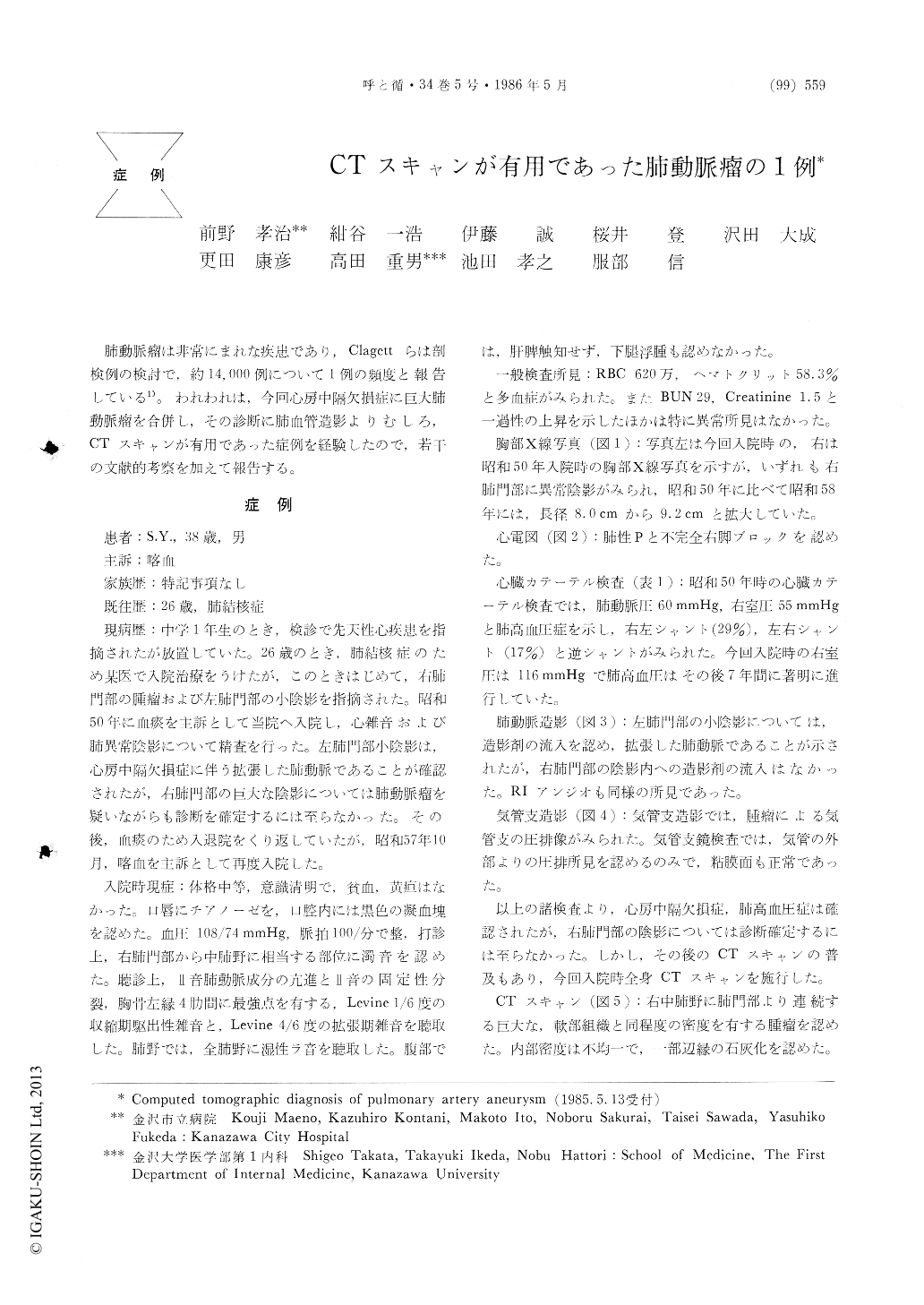
肺動脈瘤は非常にまれな疾患であり,Clagettらは剖検例の検討で,約14,000例について1例の頻度と報告している1)。われわれは,今回心房中隔欠損症に巨大肺動脈瘤を合併し,その診断に肺血管造影よりむしろ,CTスキャンが有用であった症例を経験したので,若干の文献的考察を加えて報告する。
Pulmonary artery aneurysms are rare lesions. Clagett et al reported that one aneurysm of the pulmonary artery may be found in approximately 14,000 necrop-sies. We have experienced a case of giant pulmonary artery aneurysm confirmed by computed tomography.
A 38 year-old man with atrial septal defect admitted to Kanazawa City Hospital. He had been pointed out of a right hilar mass when he was 26 years old. His complaint was bloody sputum and cough. Pulmonary angiography was not useful for the definite diagnosis because of its mural thrombi. Enhanced computed tomography showed a giant pulmonary artery aneurysm with a mural thrombi in its cavity. This shows that enhanced computed tomography is very useful for the diagnosis of a pulmonary artery aneurysm with a mural thrombi in its cavity.
Copyright © 1986, Igaku-Shoin Ltd. All rights reserved.