Japanese
English
- 有料閲覧
PPV購入案内
1,320円 (1,200円+税10%) こちらは、799ページから803ページの文献です。購入には医書.jp本会員登録が必要です。
会員登録完了後に改めて上記「購入サイトへ」を選択してください。
または「購入サイトへ」選択後に「購入ログイン」、「新規会員登録」の順に進んでください。
会員登録について詳しくはをご確認ください。
- Abstract 文献概要
- 1ページ目 Look Inside
目的
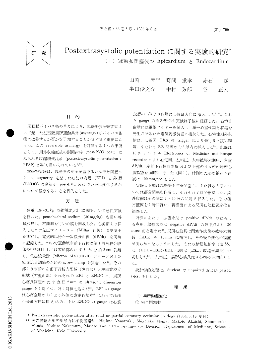
冠動脈バイパス術の普及により,冠動脈狭窄病変によって起った左室壁局所運動異常(asynergy)がバイパス術後に改善するか否かを予知することがますます重要になった。このreversible asynergyを評価する1つの手段として,期外収縮直後の洞調律時(post-PVC beat)にみられる収縮増強現象(postextrasystolic potentiation:PESP)が広く用いられている1,2)。
本動物実験は,冠動脈の完全閉塞あるいは部分閉塞によってasynergyを呈した心筋の内層(EPI)と外層(ENDO)の動態が,post-PVC beatでいかに変化するかについて観察することを目的とした。
Postextrasystolic potentiation (PESP) is widely used to detect viable but ordinary poorly contrac-ting myocardium due to coronary artery disease. PESP following coronary artery occlusion was studied serially using pairs of ultrasonic crystals to measure epicardial (EPI) and endocardial (ENDO) contractions in ischemic and nonischemic areas of left ventricle in 12 dogs.
After five minutes of total coronary occlusion (6 dogs) and thereafter, systolic expansion developed and PESP did not occur in both ENDO and EPI of the ischemic area. In contrast, in the ischemic EPI and ENDO that were akinetic, PESP persisted even after 2 hrs following partial coronary occlusion (6 dogs). PESP changed little in EPI and ENDO of the nonischemic areas after either total or partial coronary occlusion. Thus, loss of PESP occurred rapidly in not only ENDO but EN, which has different vulnerability to ischemia, following total coronaly occlusion. PESP was retained in both ENDO and EPI for at least 2 hrs following partial coronary occlusion.
Copyright © 1985, Igaku-Shoin Ltd. All rights reserved.