Japanese
English
- 有料閲覧
PPV購入案内
1,320円 (1,200円+税10%) こちらは、382ページから386ページの文献です。購入には医書.jp本会員登録が必要です。
会員登録完了後に改めて上記「購入サイトへ」を選択してください。
または「購入サイトへ」選択後に「購入ログイン」、「新規会員登録」の順に進んでください。
会員登録について詳しくはをご確認ください。
- Abstract 文献概要
- 1ページ目 Look Inside
- 参考文献 Reference
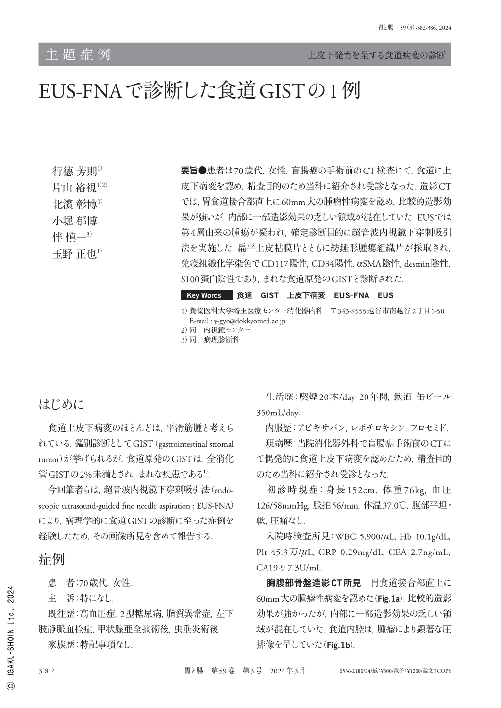
要旨●患者は70歳代,女性.盲腸癌の手術前のCT検査にて,食道に上皮下病変を認め,精査目的のため当科に紹介され受診となった.造影CTでは,胃食道接合部直上に60mm大の腫瘤性病変を認め,比較的造影効果が強いが,内部に一部造影効果の乏しい領域が混在していた.EUSでは第4層由来の腫瘍が疑われ,確定診断目的に超音波内視鏡下穿刺吸引法を実施した.扁平上皮粘膜片とともに紡錘形腫瘍組織片が採取され,免疫組織化学染色でCD117陽性,CD34陽性,αSMA陰性,desmin陰性,S100蛋白陰性であり,まれな食道原発のGISTと診断された.
A female patient in her 70s was referred to our facility for a close examination of a subepithelial esophageal tumor that was detected during a preoperative CT(computed tomography)scan for cecal cancer. Contrast-enhanced CT revealed a large 60-mm mass lesion just above the gastroesophageal junction. Some areas exhibited a relatively strong contrast effect, whereas others that were inside the mass exhibited poor contrast effect. Endoscopic ultrasonography revealed a tumor originating from the fourth layer of the esophageal wall, and endoscopic ultrasound needle aspiration was performed to confirm the diagnosis. Pathologically, the tumor was composed of the proliferation of neoplastic spindle cells that are immunohistochemically positive for CD(cluster of differentiation)117 and CD34, and negative for alpha-smooth muscle actin, desmin, and S100 proteins. Hence, the patient was diagnosed with esophageal gastrointestinal stromal tumor, which is a rare mesenchymal esophageal tumor.
Copyright © 2024, Igaku-Shoin Ltd. All rights reserved.