Japanese
English
- 有料閲覧
PPV購入案内
1,320円 (1,200円+税10%) こちらは、79ページから86ページの文献です。購入には医書.jp本会員登録が必要です。
会員登録完了後に改めて上記「購入サイトへ」を選択してください。
または「購入サイトへ」選択後に「購入ログイン」、「新規会員登録」の順に進んでください。
会員登録について詳しくはをご確認ください。
- Abstract 文献概要
- 1ページ目 Look Inside
- 参考文献 Reference
- サイト内被引用 Cited by
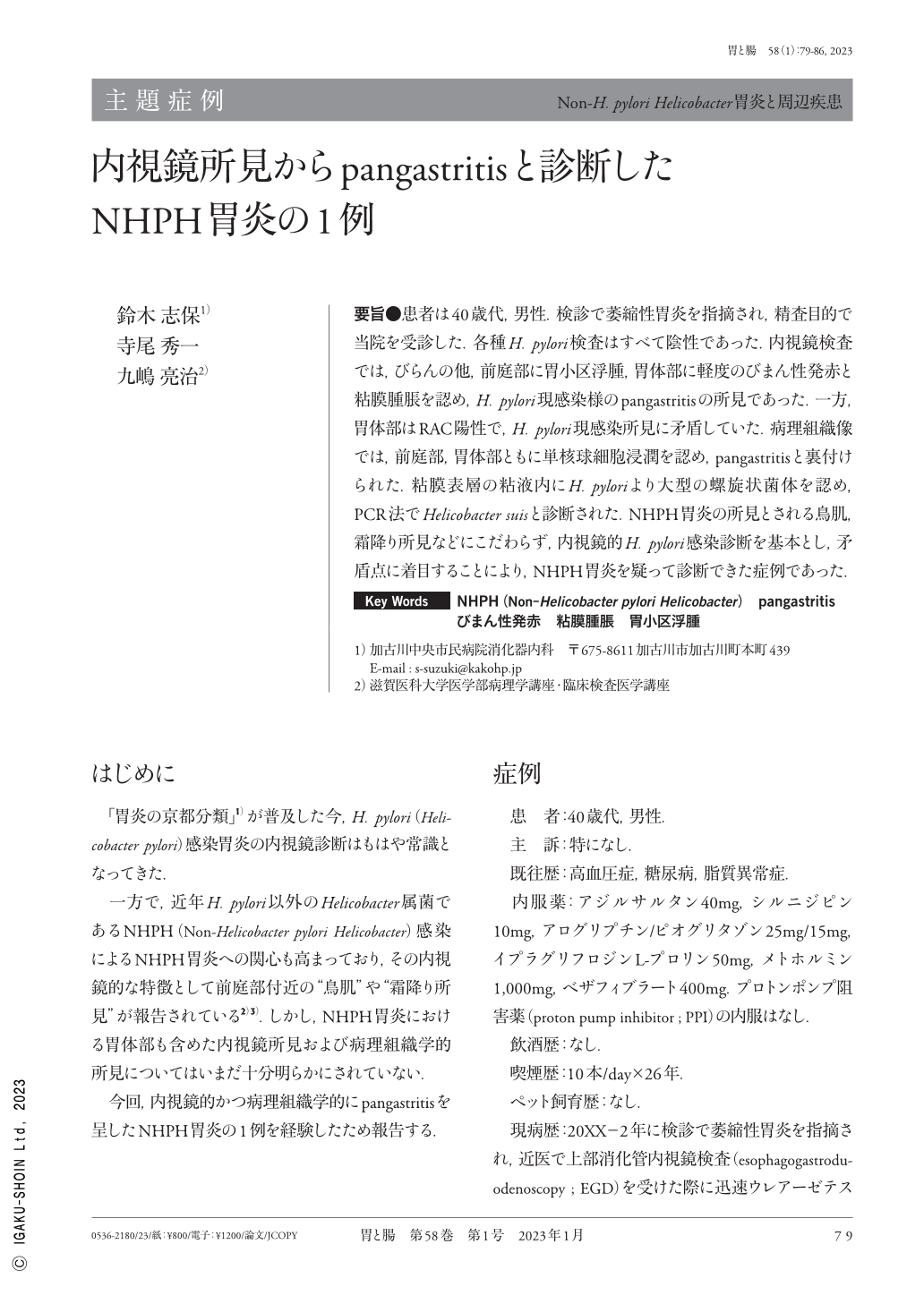
要旨●患者は40歳代,男性.検診で萎縮性胃炎を指摘され,精査目的で当院を受診した.各種H. pylori検査はすべて陰性であった.内視鏡検査では,びらんの他,前庭部に胃小区浮腫,胃体部に軽度のびまん性発赤と粘膜腫脹を認め,H. pylori現感染様のpangastritisの所見であった.一方,胃体部はRAC陽性で,H. pylori現感染所見に矛盾していた.病理組織像では,前庭部,胃体部ともに単核球細胞浸潤を認め,pangastritisと裏付けられた.粘膜表層の粘液内にH. pyloriより大型の螺旋状菌体を認め,PCR法でHelicobacter suisと診断された.NHPH胃炎の所見とされる鳥肌,霜降り所見などにこだわらず,内視鏡的H. pylori感染診断を基本とし,矛盾点に着目することにより,NHPH胃炎を疑って診断できた症例であった.
A male patient in his 40s who was diagnosed with chronic gastritis during a medical checkup visited our hospital for a thorough examination. A close upper gastrointestinal endoscopy, showed erosion and edematous areae gastricae in the gastric antrum and mild diffuse redness and mucosal edema in the gastric corpus. These findings appeared to be consistent with pangastritis caused by H. pylori(Helicobacter pylori)infection. However, it was inconsistent with H. pylori current infection because a regular arrangement of collecting venules was also observed in the gastric corpus. On the other hand, no nodular or marble appearances were observed, which have been reported in NHPH(Non-Helicobacter pylori Helicobacter)gastritis. Subsequently, all tests for H. pylori were negative. Histopathological examination revealed mononuclear cell infiltration in the gastric antrum and corpus, indicating histological pangastritis. Helical-shaped bacteria larger than H. pylori were found in the mucus collected from mucosal surface, which was identified as Helicobacter suis through a polymerase chain reaction test. In conclusion, we presented here a case led to the diagnosis of pangastritis type of NHPH by noticing the discrepancy with H. pylori and NHPH gastritis as previously reported.
Copyright © 2023, Igaku-Shoin Ltd. All rights reserved.