Japanese
English
- 有料閲覧
- Abstract 文献概要
- 1ページ目 Look Inside
- 参考文献 Reference
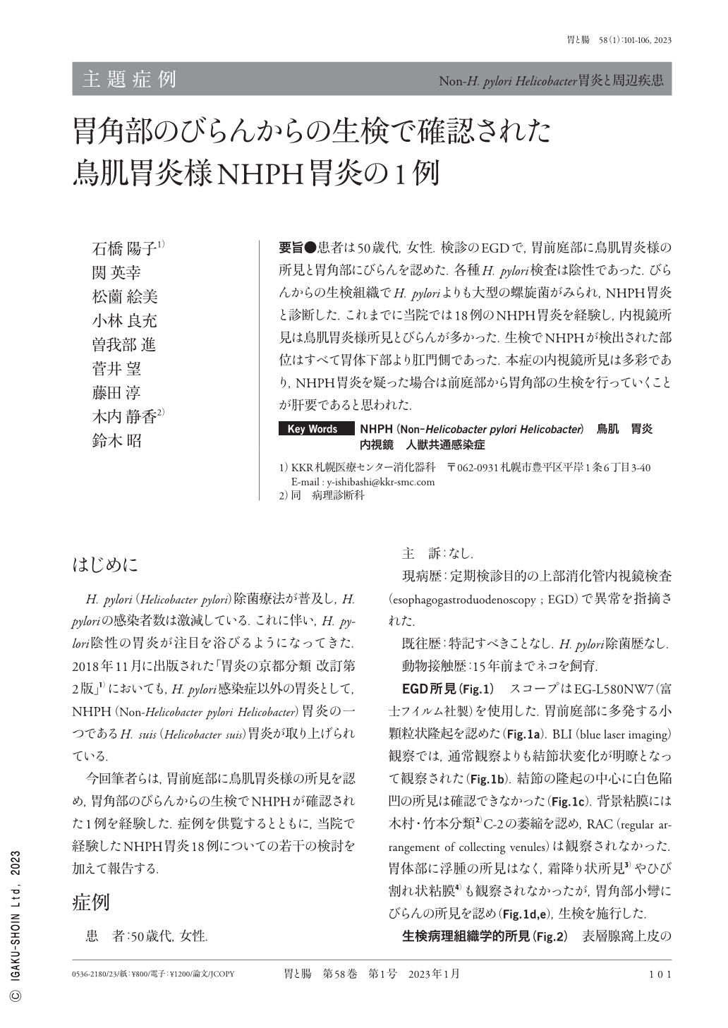
要旨●患者は50歳代,女性.検診のEGDで,胃前庭部に鳥肌胃炎様の所見と胃角部にびらんを認めた.各種H. pylori検査は陰性であった.びらんからの生検組織でH. pyloriよりも大型の螺旋菌がみられ,NHPH胃炎と診断した.これまでに当院では18例のNHPH胃炎を経験し,内視鏡所見は鳥肌胃炎様所見とびらんが多かった.生検でNHPHが検出された部位はすべて胃体下部より肛門側であった.本症の内視鏡所見は多彩であり,NHPH胃炎を疑った場合は前庭部から胃角部の生検を行っていくことが肝要であると思われた.
A woman in her 50s was referred to our institution for medical checkups without any particular symptoms. Gastrointestinal endoscopy was performed, which revealed antral nodular gastritis with erosion in the lesser curvature angle. A rapid urease test, a urea breath test, and H. pylori(Helicobacter pylori)stool antigen were all negative. A serum anti-H. pylori antibody was <3.0U/ml. Histological examination of biopsy specimens from the erosion showed more tightly coiled and longer bacteria than H. pylori, suggesting a NHPH(Non-Helicobacter pylori Helicobacter)infection. For 11 years, we have encountered a total of 18 cases of NHPH gastritis. NHPH was detected in the lesion from the lower body to the antrum, but not from the middle to the upper body, in all cases. Due to its various characteristics, details of endoscopic findings of NHPH gastritis have not been clarified. Therefore, endoscopists should consider this rare entity as a differential diagnosis and obtain biopsy specimens from the antrum or angle of the stomach.

Copyright © 2023, Igaku-Shoin Ltd. All rights reserved.


