Japanese
English
- 有料閲覧
- Abstract 文献概要
- 1ページ目 Look Inside
- 参考文献 Reference
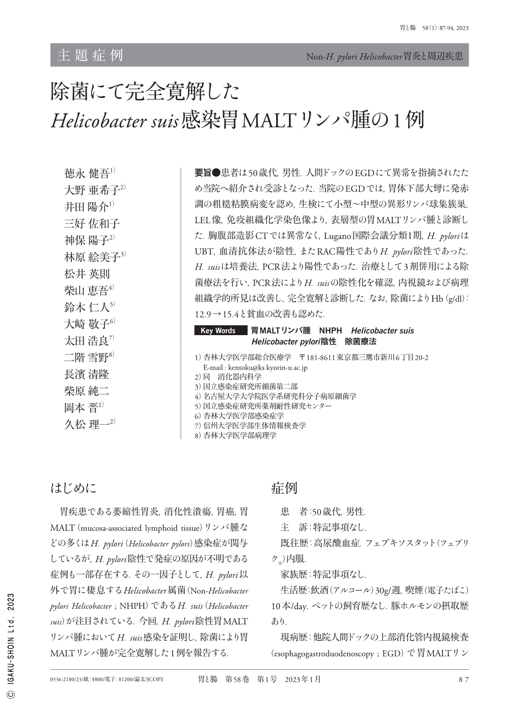
要旨●患者は50歳代,男性.人間ドックのEGDにて異常を指摘されたため当院へ紹介され受診となった.当院のEGDでは,胃体下部大彎に発赤調の粗糙粘膜病変を認め,生検にて小型〜中型の異形リンパ球集簇巣,LEL像,免疫組織化学染色像より,表層型の胃MALTリンパ腫と診断した.胸腹部造影CTでは異常なく,Lugano国際会議分類I期,H. pyloriはUBT,血清抗体法が陰性,またRAC陽性でありH. pylori陰性であった.H. suisは培養法,PCR法より陽性であった.治療として3剤併用による除菌療法を行い,PCR法によりH. suisの陰性化を確認,内視鏡および病理組織学的所見は改善し,完全寛解と診断した.なお,除菌によりHb(g/dl):12.9→15.4と貧血の改善も認めた.
A man in his 50s underwent upper gastrointestinal endoscopy due to an abnormality observed during a medical checkup. The EGD(esophagogastroduodenoscopy)revealed reddish rough mucosa along the greater curvature of the lower body of the stomach. Based on the pathological findings, like atypical lymphocyte clusters, lymphoepithelial lesion images, and immunostaining images, a diagnosis of gastric MALT(mucosa-associated lymphoid tissue)lymphoma was made. The urea breath test and serum antibody tests were negative for Helicobacter pylori infection. However, the culture and polymerase chain reaction methods were positive for H. suis(Helicobacter suis)infection. Hence, H. suis-infected gastric MALT lymphoma of Lugano stage I was diagnosed. Eradication therapy with triple-drug therapy was prescribed, and H. suis-negative conversion was confirmed. The EGD and pathological findings showed an improvement. Also, the anemia improved after initiating the eradication therapy.

Copyright © 2023, Igaku-Shoin Ltd. All rights reserved.


