Japanese
English
- 有料閲覧
PPV購入案内
1,320円 (1,200円+税10%) こちらは、1083ページから1091ページの文献です。購入には医書.jp本会員登録が必要です。
会員登録完了後に改めて上記「購入サイトへ」を選択してください。
または「購入サイトへ」選択後に「購入ログイン」、「新規会員登録」の順に進んでください。
会員登録について詳しくはをご確認ください。
- Abstract 文献概要
- 1ページ目 Look Inside
- 参考文献 Reference
- サイト内被引用 Cited by
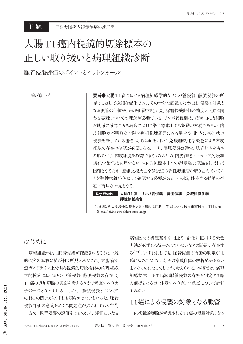
要旨●大腸T1癌における病理組織学的なリンパ管侵襲,静脈侵襲の所見はしばしば微細な変化であり,その十分な認識のためには,侵襲の対象となる脈管の部位や,病理組織学的所見,脈管侵襲評価の精度と限界に関わる要因についての理解が必要である.リンパ管侵襲は,腔縁に内皮細胞が明確に確認できる場合にはHE染色標本上でも認識が容易であるが,内皮細胞が不明瞭な空隙を癌細胞塊周囲にみる場合や,腔内に塞栓状の侵襲を来している場合は,D2-40を用いた免疫組織化学染色による内皮細胞の存在の確認が必要となる.一方,静脈侵襲は通常,脈管腔内を占める形で生じ,内皮細胞を確認できなくなるため,内皮細胞マーカーの免疫組織化学染色は有用でない.HE染色標本上での静脈壁の認識もしばしば困難となるため,癌細胞塊周囲を静脈壁の弾性線維層が取り囲んでいることを弾性線維染色により確認する必要がある.その際,伴走する動脈の存在は有用な所見となる.
Lymphovascular and venous invasions are recognized as subtle changes in the histopathologic examination of T1 colorectal cancers. Therefore, it is a prerequisite to understand the site and histologic findings of the involved vessels and the factors determining the quality and limitation of their detection. Lymphovascular invasion is easily determined when a cancer cell nest is observed in the lumen lined by unequivocal endothelial cells. However, endothelial lining is often indistinct, thereby requiring D2-40 immunohistochemistry for confirmation of the lymphovascular invasion. Venous invasion is generally observed as a cancer cell nest occupying the venous lumen with endothelial impairment, due to which the studies on immunohistochemical endothelial markers are of no use. To clearly determine the veins involved in cancer invasion, it is necessary to incorporate elastic fiber stain that helps in demonstrating the elastic fiber layers of the venous wall around the cancer cell nest. The presence of an accompanying artery provides another marker for venous invasion.
Copyright © 2021, Igaku-Shoin Ltd. All rights reserved.