Japanese
English
- 有料閲覧
PPV購入案内
1,320円 (1,200円+税10%) こちらは、1057ページから1063ページの文献です。購入には医書.jp本会員登録が必要です。
会員登録完了後に改めて上記「購入サイトへ」を選択してください。
または「購入サイトへ」選択後に「購入ログイン」、「新規会員登録」の順に進んでください。
会員登録について詳しくはをご確認ください。
- Abstract 文献概要
- 1ページ目 Look Inside
- 参考文献 Reference
- サイト内被引用 Cited by
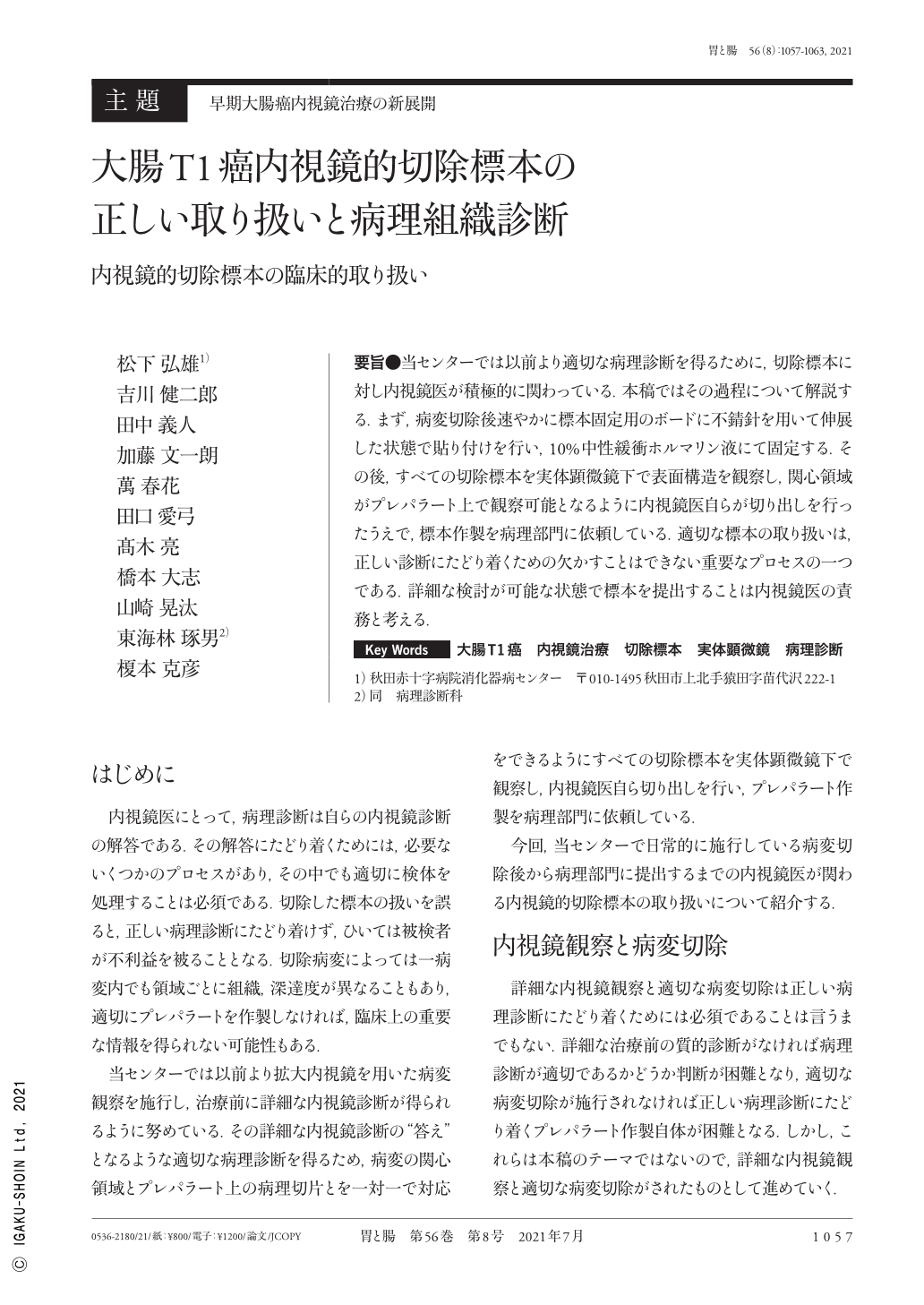
要旨●当センターでは以前より適切な病理診断を得るために,切除標本に対し内視鏡医が積極的に関わっている.本稿ではその過程について解説する.まず,病変切除後速やかに標本固定用のボードに不錆針を用いて伸展した状態で貼り付けを行い,10%中性緩衝ホルマリン液にて固定する.その後,すべての切除標本を実体顕微鏡下で表面構造を観察し,関心領域がプレパラート上で観察可能となるように内視鏡医自らが切り出しを行ったうえで,標本作製を病理部門に依頼している.適切な標本の取り扱いは,正しい診断にたどり着くための欠かすことはできない重要なプロセスの一つである.詳細な検討が可能な状態で標本を提出することは内視鏡医の責務と考える.
The resected specimens of colorectal T1 cancer are appropriately treated to obtain an accurate pathological diagnosis. Immediately after the lesion excision, the lesion is attached to the specimen fixing board using a pin. Then, it is fixed with 10% neutral buffered formalin solution. Then, the surface structure of all excised specimens is observed in detail under a stereomicroscope. An endoscopist makes incisions so that the area of interest is a pathological specimen. Thereafter, the resected specimen is submitted to the pathology department.
Proper handling of resected specimens is one of the important processes that facilitate accurate diagnosis. It is the responsibility of the endoscopist to submit the specimen in a state where detailed examination is possible.
Copyright © 2021, Igaku-Shoin Ltd. All rights reserved.