Japanese
English
- 有料閲覧
PPV購入案内
1,320円 (1,200円+税10%) こちらは、1131ページから1148ページの文献です。購入には医書.jp本会員登録が必要です。
会員登録完了後に改めて上記「購入サイトへ」を選択してください。
または「購入サイトへ」選択後に「購入ログイン」、「新規会員登録」の順に進んでください。
会員登録について詳しくはをご確認ください。
- Abstract 文献概要
- 1ページ目 Look Inside
- 参考文献 Reference
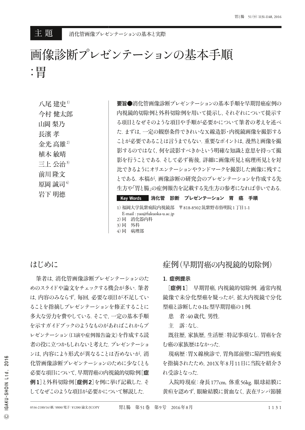
要旨●消化管画像診断プレゼンテーションの基本手順を早期胃癌症例の内視鏡的切除例と外科切除例を用いて提示し,それぞれについて提示する項目となぜそのような項目や手順が必要かについて筆者の考えを述べた.まずは,一定の観察条件できれいなX線造影・内視鏡画像を撮影することが必要であることは言うまでもない.重要なポイントは,漫然と画像を撮影するのではなく,何を読影すべきかという明確な知識と意思を持って撮影を行うことである.そして必ず術後,詳細に画像所見と病理所見とを対比できるようにオリエンテーションやランドマークを撮影した画像に残すことである.本稿が,画像診断の研究会のプレゼンテーションを作成する先生方や「胃と腸」の症例報告を記載する先生方の参考になれば幸いである.
Here, we demonstrated how to create a presentation for gastrointestinal imaging using two representative cases with early gastric cancers, one involving endoscopic resection and the other involving surgical resection. Furthermore, we addressed why the involvement of each subject was mandatory for the presentation. It is very important to obtain an excellent radiological and endoscopic image under a specific condition. During procedures, doctors therefore need to consider why such images are important for arriving at correct and precise diagnoses. To accurately make comparisons between images and histological architecture following resection, it is preferable to identify a landmark on the images themselves or to place an identification mark alongside the target lesion. We sincerely hope that this manuscript adequately describes a minimal required standard which could become useful for all doctors attempting to create good quality presentations for gastrointestinal imaging diagnosis.
Copyright © 2016, Igaku-Shoin Ltd. All rights reserved.