Japanese
English
- 有料閲覧
PPV購入案内
1,320円 (1,200円+税10%) こちらは、1749ページから1753ページの文献です。購入には医書.jp本会員登録が必要です。
会員登録完了後に改めて上記「購入サイトへ」を選択してください。
または「購入サイトへ」選択後に「購入ログイン」、「新規会員登録」の順に進んでください。
会員登録について詳しくはをご確認ください。
- Abstract 文献概要
- 1ページ目 Look Inside
- 参考文献 Reference
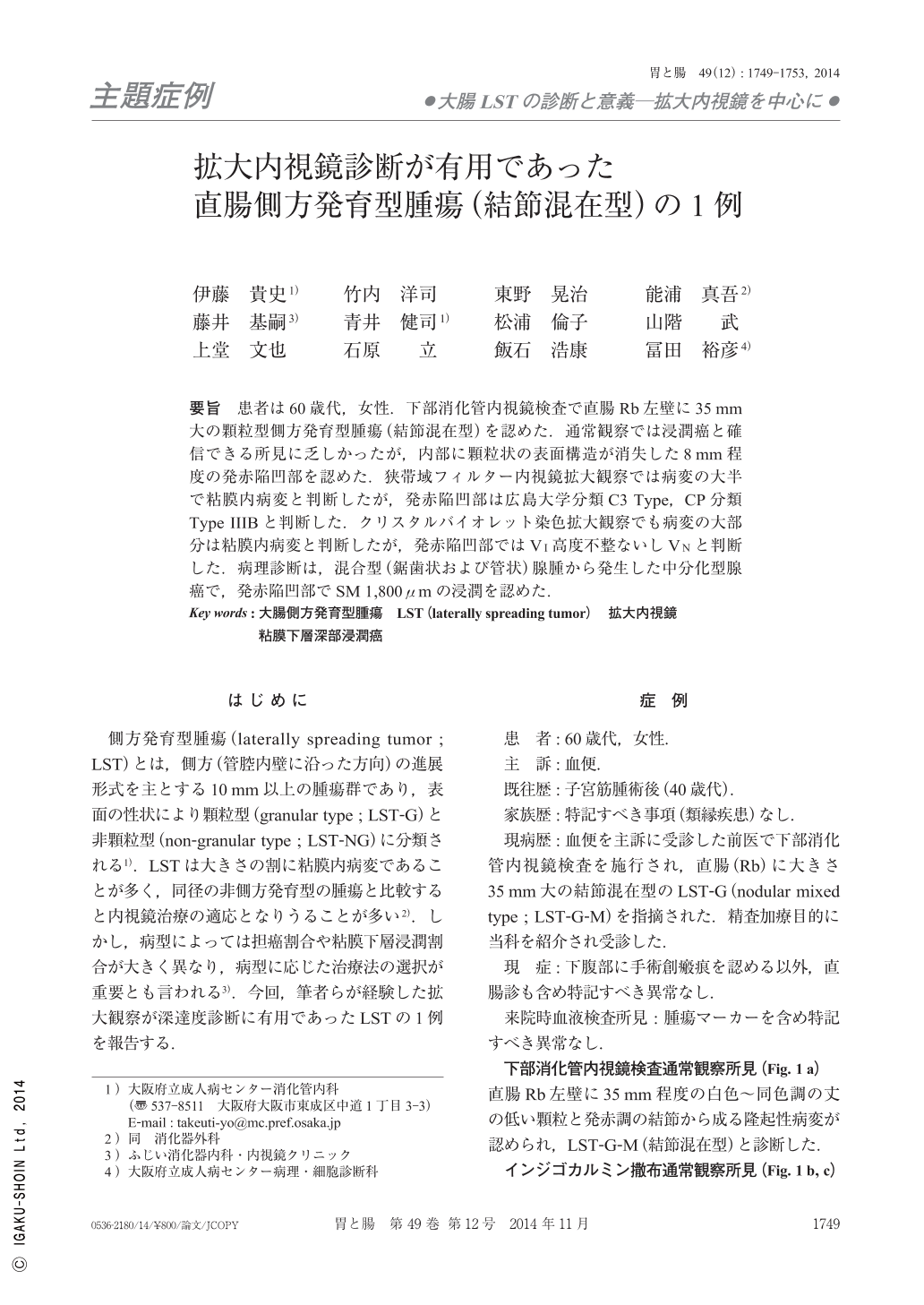
要旨 患者は60歳代,女性.下部消化管内視鏡検査で直腸Rb左壁に35mm大の顆粒型側方発育型腫瘍(結節混在型)を認めた.通常観察では浸潤癌と確信できる所見に乏しかったが,内部に顆粒状の表面構造が消失した8mm程度の発赤陥凹部を認めた.狭帯域フィルター内視鏡拡大観察では病変の大半で粘膜内病変と判断したが,発赤陥凹部は広島大学分類C3 Type,CP分類Type IIIBと判断した.クリスタルバイオレット染色拡大観察でも病変の大部分は粘膜内病変と判断したが,発赤陥凹部ではVI高度不整ないしVNと判断した.病理診断は,混合型(鋸歯状および管状)腺腫から発生した中分化型腺癌で,発赤陥凹部でSM 1,800μmの浸潤を認めた.
A woman in her sixties was referred to our hospital for follow-up for a lesion detected in the colon in another hospital. A colonoscopy showed a 35mm-sized granular-type laterally spreading tumor(nodular-mixed type)on the left wall of the rectum. Although no findings suggested invasion into the submucosal layer, we noticed a reddish and depressed area where the granular surface disappeared on conventional endoscopy. Using magnifying narrow-band imaging, majority of the tumor was diagnosed as an intramucosal lesion, whereas the reddish and depressed area was diagnosed as C3 type in the Hiroshima University classification and type IIIB in the Capillary pattern classification. In addition, most of the lesion was also suspected as an intramucosal lesion by pit pattern diagnosis with crystal violet dye ; however, the pit pattern of the reddish and depressed area was diagnosed as VI or VN. Therefore, we speculated that the lesion was a carcinoma deeply invaded into the submucosal layer at the reddish and depressed area. Histological findings of the resected specimen showed that the tumor was a moderately-differentiated adenocarcinoma that originated from a tubular and serrated adenoma. The tumor invaded into the submucosa deeply(1,800μm)at the reddish and depressed area.
Copyright © 2014, Igaku-Shoin Ltd. All rights reserved.