Japanese
English
- 有料閲覧
PPV購入案内
1,320円 (1,200円+税10%) こちらは、1141ページから1156ページの文献です。購入には医書.jp本会員登録が必要です。
会員登録完了後に改めて上記「購入サイトへ」を選択してください。
または「購入サイトへ」選択後に「購入ログイン」、「新規会員登録」の順に進んでください。
会員登録について詳しくはをご確認ください。
- Abstract 文献概要
- 1ページ目 Look Inside
- 参考文献 Reference
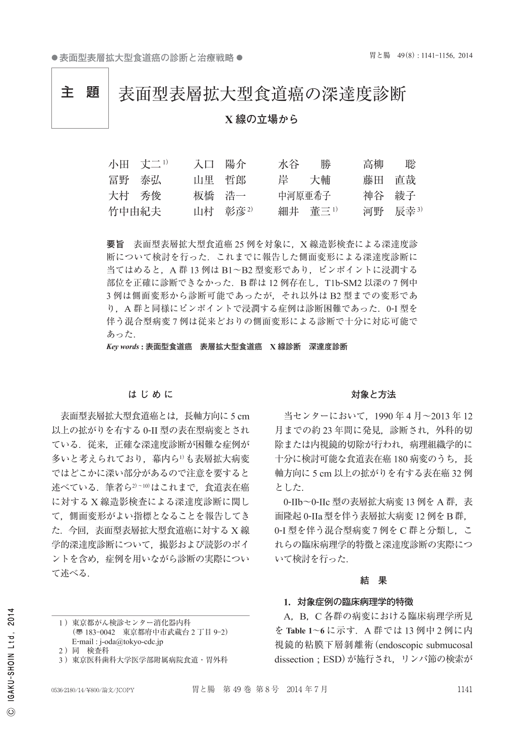
要旨 表面型表層拡大型食道癌25例を対象に,X線造影検査による深達度診断について検討を行った.これまでに報告した側面変形による深達度診断に当てはめると,A群13例はB1~B2型変形であり,ピンポイントに浸潤する部位を正確に診断できなかった.B群は12例存在し,T1b-SM2以深の7例中3例は側面変形から診断可能であったが,それ以外はB2型までの変形であり,A群と同様にピンポイントで浸潤する症例は診断困難であった.0-I型を伴う混合型病変7例は従来どおりの側面変形による診断で十分に対応可能であった.
The present study investigated the radiological diagnosis of the depth of invasion in 25 cases of superficial spreading type of esophageal carcinoma. Thirteen cases in Group A showed type B1 or B2 deformation on the lateral view, but the area of spread could not be precisely identified. Group B included 12 cases, and the depth of invasion could be determined based on deformation on the lateral view in 3 of 7 cases of carcinoma in the deep sub-mucosal layer. However, for Group A, where the area of spread could not be identified, an assessment of depth was difficult. Overall, it was possible to diagnose the depth of invasion in 7 cases with stage 0 or I cancer based on deformation on the lateral view precisely as before.
Copyright © 2014, Igaku-Shoin Ltd. All rights reserved.