Japanese
English
- 有料閲覧
- Abstract 文献概要
- 1ページ目 Look Inside
- 参考文献 Reference
- サイト内被引用 Cited by
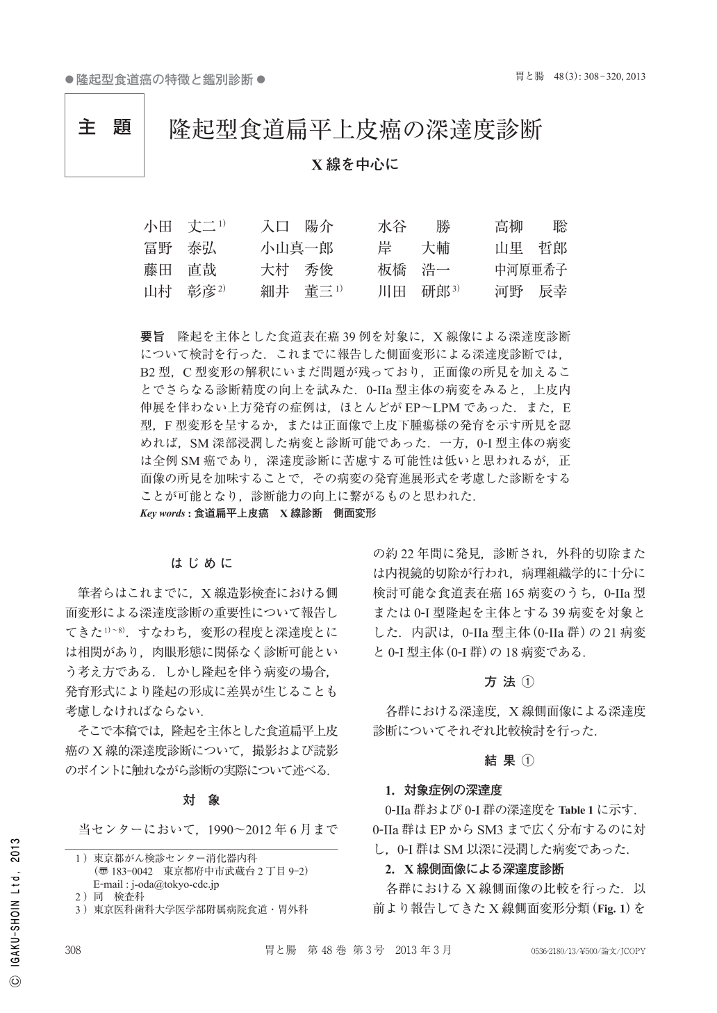
要旨 隆起を主体とした食道表在癌39例を対象に,X線像による深達度診断について検討を行った.これまでに報告した側面変形による深達度診断では,B2型,C型変形の解釈にいまだ問題が残っており,正面像の所見を加えることでさらなる診断精度の向上を試みた.0-IIa型主体の病変をみると,上皮内伸展を伴わない上方発育の症例は,ほとんどがEP~LPMであった.また,E型,F型変形を呈するか,または正面像で上皮下腫瘍様の発育を示す所見を認めれば,SM深部浸潤した病変と診断可能であった.一方,0-I型主体の病変は全例SM癌であり,深達度診断に苦慮する可能性は低いと思われるが,正面像の所見を加味することで,その病変の発育進展形式を考慮した診断をすることが可能となり,診断能力の向上に繋がるものと思われた.
We investigated about 39 superficial carcinomas of the esophagus by radiological diagnosis of depth of invasion. In radiological diagnosis of the depth of invasion from deformation of the lateral view, the problem still remains in the interpretation of B2 type and C type deformation. So, We tried improvement in diagnostic accuracy by adding the radiological view of the front image. Most of the cases of 0-IIa type, with upward-growth without intraepithelial spread were EP-LPM. When recognizing E or F type deformation of the lateral view or subepithelial growth by the radiological front image, diagnosis of the depth of invasion was possible for the deep sub-mucosal layer. On the other hand, all cases of 0-I type invaded to the sub-mucosal layer, so diagnosis for the depth of invasion was not so difficult. I think we can diagnose in consideration of the growth progression, and improve our diagnostic capability by adding the radiological view of the front image.

Copyright © 2013, Igaku-Shoin Ltd. All rights reserved.


