Japanese
English
- 有料閲覧
PPV購入案内
1,320円 (1,200円+税10%) こちらは、137ページから147ページの文献です。購入には医書.jp本会員登録が必要です。
会員登録完了後に改めて上記「購入サイトへ」を選択してください。
または「購入サイトへ」選択後に「購入ログイン」、「新規会員登録」の順に進んでください。
会員登録について詳しくはをご確認ください。
- Abstract 文献概要
- 1ページ目 Look Inside
- 参考文献 Reference
- サイト内被引用 Cited by
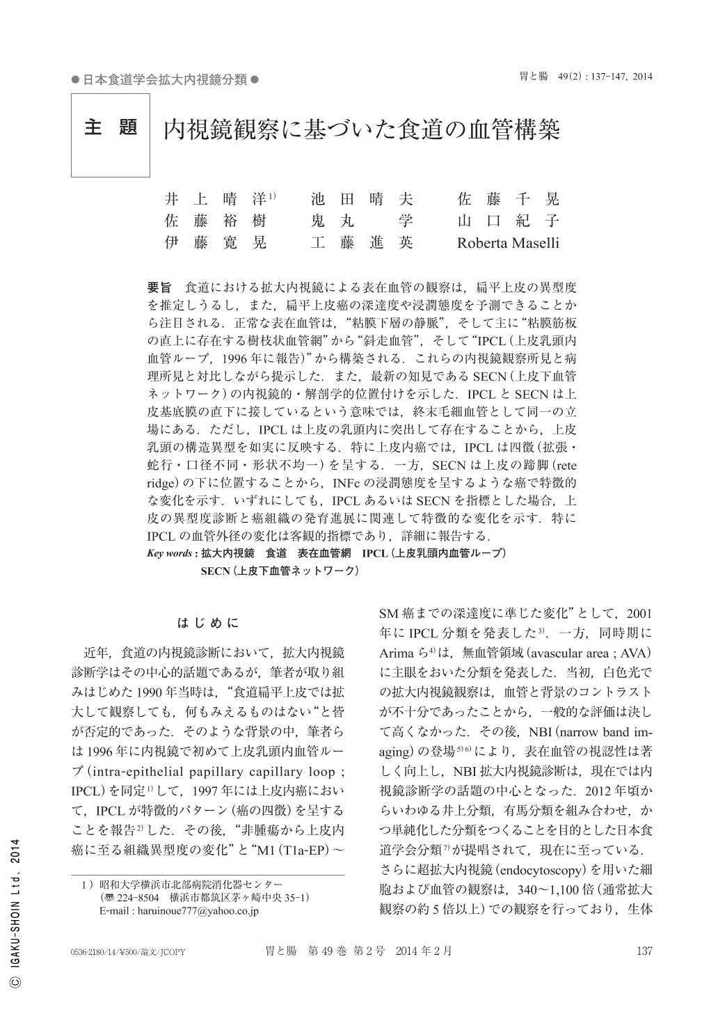
要旨 食道における拡大内視鏡による表在血管の観察は,扁平上皮の異型度を推定しうるし,また,扁平上皮癌の深達度や浸潤態度を予測できることから注目される.正常な表在血管は,“粘膜下層の静脈”,そして主に“粘膜筋板の直上に存在する樹枝状血管網”から“斜走血管”,そして“IPCL(上皮乳頭内血管ループ,1996年に報告)”から構築される.これらの内視鏡観察所見と病理所見と対比しながら提示した.また,最新の知見であるSECN(上皮下血管ネットワーク)の内視鏡的・解剖学的位置付けを示した.IPCLとSECNは上皮基底膜の直下に接しているという意味では,終末毛細血管として同一の立場にある.ただし,IPCLは上皮の乳頭内に突出して存在することから,上皮乳頭の構造異型を如実に反映する.特に上皮内癌では,IPCLは四徴(拡張・蛇行・口径不同・形状不均一)を呈する.一方,SECNは上皮の蹄脚(rete ridge)の下に位置することから,INFcの浸潤態度を呈するような癌で特徴的な変化を示す.いずれにしても,IPCLあるいはSECNを指標とした場合,上皮の異型度診断と癌組織の発育進展に関連して特徴的な変化を示す.特にIPCLの血管外径の変化は客観的指標であり,詳細に報告する.
Recent advancement of endoscopic imaging technology(NBI magnifying endoscopy)enables precise observation of superficial vascular structure in the esophagus. Superficial vasculature in the esophagus consists of“submucosal vein”,“branching vein network mainly just above muscularis mucosa”,“oblique running vessel”and“IPCL(intra-epithelial papillary capillary loop)”. Endoscopic findings of these vessels were firstly reported in 1996. Among them, IPCL is positioned at epithelium papilla and demonstrates various figure changes according to increase of tissue atypia. In carcinoma in situ, IPCL shows 4 characteristic figure changes ; dilation, meandering, irregular caliber, and shape variation. Recently, SECN(subepithelial capillary network)was also identified as a tiny capillary network just beneath epithelium. SECN changes closely reflects diffuse invasion(INFc)of cancer tissue. In the squamous cell carcinoma, IPCL demonstrates typical figure changes according to cancer invasion depth and finally destroyed and totally disappeared replaced by new tumor vessels. When talking about cancer invasion depth, abnormal vessel size is most objective findings. Normal IPCL has 7.7μm diameter, and B1(IPCL type V-1)demonstrates 21.9μm in size, and B3(IPCL type VN)shows 67.2μm on average. In this article, this process is precisely reported contrasting with histological findings.
Copyright © 2014, Igaku-Shoin Ltd. All rights reserved.