Japanese
English
- 有料閲覧
PPV購入案内
1,320円 (1,200円+税10%) こちらは、564ページから572ページの文献です。購入には医書.jp本会員登録が必要です。
会員登録完了後に改めて上記「購入サイトへ」を選択してください。
または「購入サイトへ」選択後に「購入ログイン」、「新規会員登録」の順に進んでください。
会員登録について詳しくはをご確認ください。
- Abstract 文献概要
- 1ページ目 Look Inside
- 参考文献 Reference
- サイト内被引用 Cited by
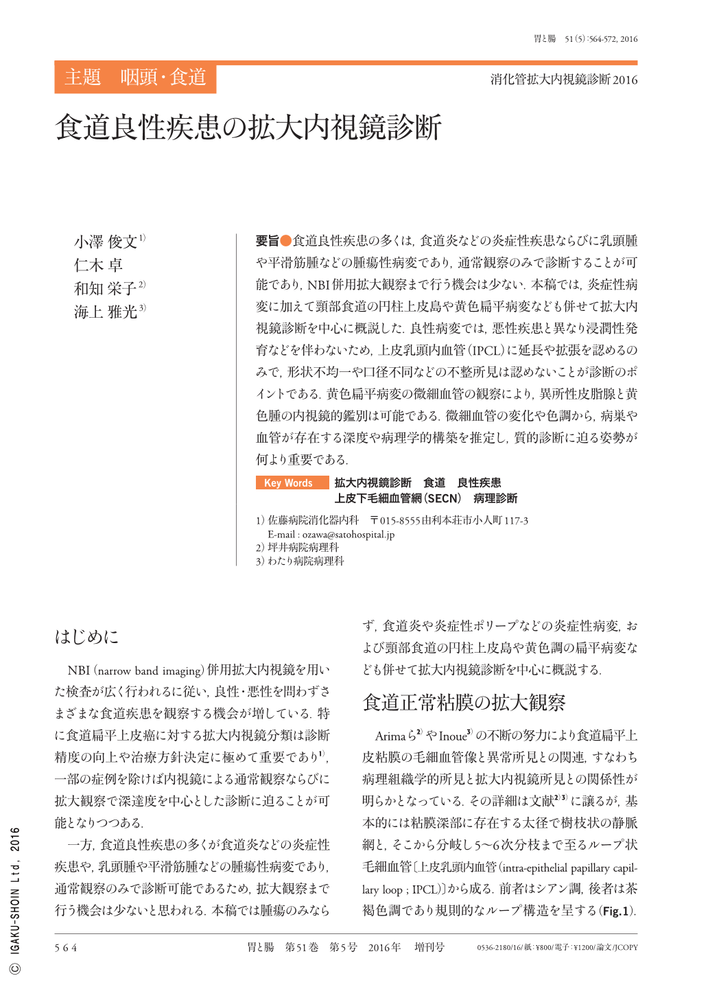
要旨●食道良性疾患の多くは,食道炎などの炎症性疾患ならびに乳頭腫や平滑筋腫などの腫瘍性病変であり,通常観察のみで診断することが可能であり,NBI併用拡大観察まで行う機会は少ない.本稿では,炎症性病変に加えて頸部食道の円柱上皮島や黄色扁平病変なども併せて拡大内視鏡診断を中心に概説した.良性病変では,悪性疾患と異なり浸潤性発育などを伴わないため,上皮乳頭内血管(IPCL)に延長や拡張を認めるのみで,形状不均一や口径不同などの不整所見は認めないことが診断のポイントである.黄色扁平病変の微細血管の観察により,異所性皮脂腺と黄色腫の内視鏡的鑑別は可能である.微細血管の変化や色調から,病巣や血管が存在する深度や病理学的構築を推定し,質的診断に迫る姿勢が何より重要である.
Almost all benign esophageal diseases are classified as inflammatory reflux diseases, or neoplastic diseases represented by papillomas or leiomyomas. These disorders are easily diagnosed using ordinary white light endoscopy.
The aim of this report was to investigate the correlation of the magnifying endoscopic findings, using narrow band imaging, and histological findings of benign esophageal lesions, containing ectopic gastric mucosa(cervical)and flat-yellowish irregularities. We postulated that benign esophageal lesions are often accompanied by mild changes of the intra-papillary capillary loop including elongation and/or dilation with no irregularity. We concluded that a differential diagnosis of ectopic sebaceous glands and xanthoma in the esophagus was possible by magnifying endoscopy.
Therefore, we recommend careful examination and analysis of correlations between endoscopic microvascular and histological findings of various lesions.
Copyright © 2016, Igaku-Shoin Ltd. All rights reserved.