Japanese
English
- 有料閲覧
PPV購入案内
1,320円 (1,200円+税10%) こちらは、217ページから226ページの文献です。購入には医書.jp本会員登録が必要です。
会員登録完了後に改めて上記「購入サイトへ」を選択してください。
または「購入サイトへ」選択後に「購入ログイン」、「新規会員登録」の順に進んでください。
会員登録について詳しくはをご確認ください。
- Abstract 文献概要
- 1ページ目 Look Inside
- 参考文献 Reference
- サイト内被引用 Cited by
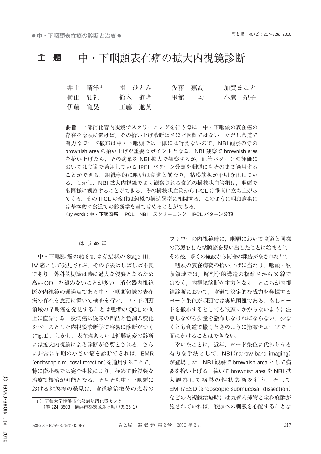
要旨 上部消化管内視鏡でスクリーニングを行う際に,中・下咽頭の表在癌の存在を念頭に置けば,その拾い上げ診断はさほど困難ではない.ただし食道で有力なヨード撒布は中・下咽頭では一律には行えないので,NBI観察の際のbrownish areaの拾い上げが重要なポイントとなる.NBI観察でbrownish areaを拾い上げたら,その病巣をNBI拡大で観察するが,血管パターンの評価においては食道で適用しているIPCLパターン分類を咽頭にもそのまま適用することができる.組織学的に咽頭は食道と異なり,粘膜筋板が不明瞭化している.しかし,NBI拡大内視鏡でよく観察される食道の樹枝状血管網は,咽頭でも同様に観察することができる.その樹枝状血管からIPCLは垂直に立ち上がってくる.そのIPCLの変化は組織の構造異型に相関する.このように咽頭病巣には基本的に食道での診断学を当てはめることができる.
When we perform endoscopic screening in the upper GI tract, superficial cancer in the middle and hypopharynx can also be detected. It is impossible to spray iodine solution in the pharynx because of its irritating effect on the larynx, and therefore NBI(narrow band imaging)image-enahanced endoscopy becomes a key tool for identifying flat lesions in the pharynx. After detecting a brownish area in the pharynx, NBI magnifying observation facilitates the evaluation of IPCL(intraepithelial papillary capillary loop)pattern changes. Histologically, lesions in the pharynx muscularis mucosae become hard to identify, but normal superficial vascular network pattern in the pharynx is generally similar to that in the esophagus. In order to make a diagnosis of superficial lesions in the pharynx, IPCL pattern classification in the esophagus can be applied also to pharyngeal lesions.
Copyright © 2010, Igaku-Shoin Ltd. All rights reserved.