Japanese
English
- 有料閲覧
PPV購入案内
1,320円 (1,200円+税10%) こちらは、337ページから348ページの文献です。購入には医書.jp本会員登録が必要です。
会員登録完了後に改めて上記「購入サイトへ」を選択してください。
または「購入サイトへ」選択後に「購入ログイン」、「新規会員登録」の順に進んでください。
会員登録について詳しくはをご確認ください。
- Abstract 文献概要
- 1ページ目 Look Inside
- 参考文献 Reference
- サイト内被引用 Cited by
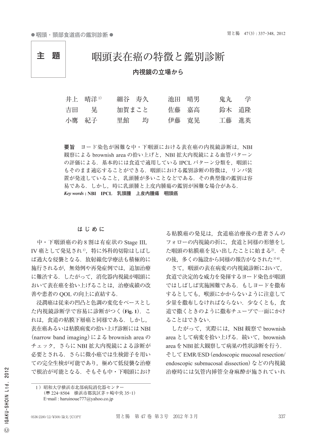
要旨 ヨード染色が困難な中・下咽頭における表在癌の内視鏡診断は,NBI観察によるbrownish areaの拾い上げと,NBI拡大内視鏡による血管パターンの評価による.基本的には食道で適用しているIPCLパターン分類を,咽頭にもそのまま適応することができる.咽頭における鑑別診断の特徴は,リンパ装置が発達していること,乳頭腫が多いことなどである.その典型像の鑑別は容易である.しかし,時に乳頭腫と上皮内腫瘍の鑑別が困難な場合がある.
Superficial cancer in the middle and hypopharynx can be detected by careful observation. It is impossible to spray iodine solution in the pharynx because it irritates the larynx, so NBI(narrow band imaging)becomes the key tool to identify flat lesions in the pharynx. After detecting a brownish area in the pharynx, NBI magnifying observation facilitates the evaluation of IPCL(intra-epithelial papillary capillary loop)pattern changes. An importanat differential diagnosis in the pharynx is squamous cell papilloma. Papillomatous lesions are easily diagnosed as squamous cell papilloma by typical endoscopic findings, but in flat lesions endoscopic diagnosis often becomes difficult.
Copyright © 2012, Igaku-Shoin Ltd. All rights reserved.