Japanese
English
- 有料閲覧
- Abstract 文献概要
- 1ページ目 Look Inside
- 参考文献 Reference
- サイト内被引用 Cited by
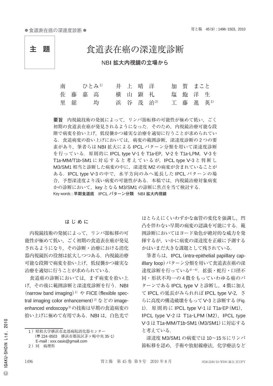
要旨 内視鏡技術の発展によって,リンパ節転移の可能性が極めて低い,ごく初期の食道表在癌が発見されるようになった.そのため,内視鏡治療可能な段階で病変を拾い上げ,低侵襲かつ確実な治療を適切に行うことが求められている.食道病変の拾い上げにおいては,病変の範囲診断,深達度診断の2つの要素があり,筆者らはNBI拡大によるIPCLパターン分類を用いて深達度診断を行っている.原則的にIPCL type V-1をT1a-EP,V-2をT1a-LPM,V-3をT1a-MM/T1b-SM1に対応すると考えているが,IPCL type V-3と判断しM3/SM1相当と診断した病変の中に,深達度M2の病変が含まれていることがある.IPCL type V-3の中で,水平方向のみへ延長したIPCLパターンの場合,予想深達度より浅い病変の可能性がある.本稿では,内視鏡治療対象病変かの診断において,keyとなるM3/SM1の診断に焦点を当て検討する.
With the outstanding development of endoscopic technology, a number of early stage esophageal cancers have been found. Nowadays, both recognizing the lesions which can still be treated by endoscopic procedures and performing safe and precise resection are essential skills for Endoscopists.
Esophageal diagnosis consists of 2 steps. The first step is to detect the lesion as a brownish area or non-iodine-stained lesion. The next step is to observe the suspected area with high magnification and then evaluate the IPCL pattern. Basically,IPCL type V-1 is thought to correspond to T1a-EP,V-2 corresponds to T1a-LPM and V-3 to T1a-MM/T1b-SM1. However, there are some cases which we diagnosed as IPCL type V-3 before resection, but revealed T1a-LPM pathologically. Definition of IPCL type V-3 is “Advanced destruction of IPCL". To diagnose the V-3 lesions more precisely, we categorized IPCL type V-3 into three subgroups and evaluated each pattern. IPCL type V-3 alone with horizontally prolonged vessels includes T1a-EP/LPM lesions.

Copyright © 2010, Igaku-Shoin Ltd. All rights reserved.


