Japanese
English
- 有料閲覧
PPV購入案内
1,320円 (1,200円+税10%) こちらは、1235ページから1239ページの文献です。購入には医書.jp本会員登録が必要です。
会員登録完了後に改めて上記「購入サイトへ」を選択してください。
または「購入サイトへ」選択後に「購入ログイン」、「新規会員登録」の順に進んでください。
会員登録について詳しくはをご確認ください。
- Abstract 文献概要
- 1ページ目 Look Inside
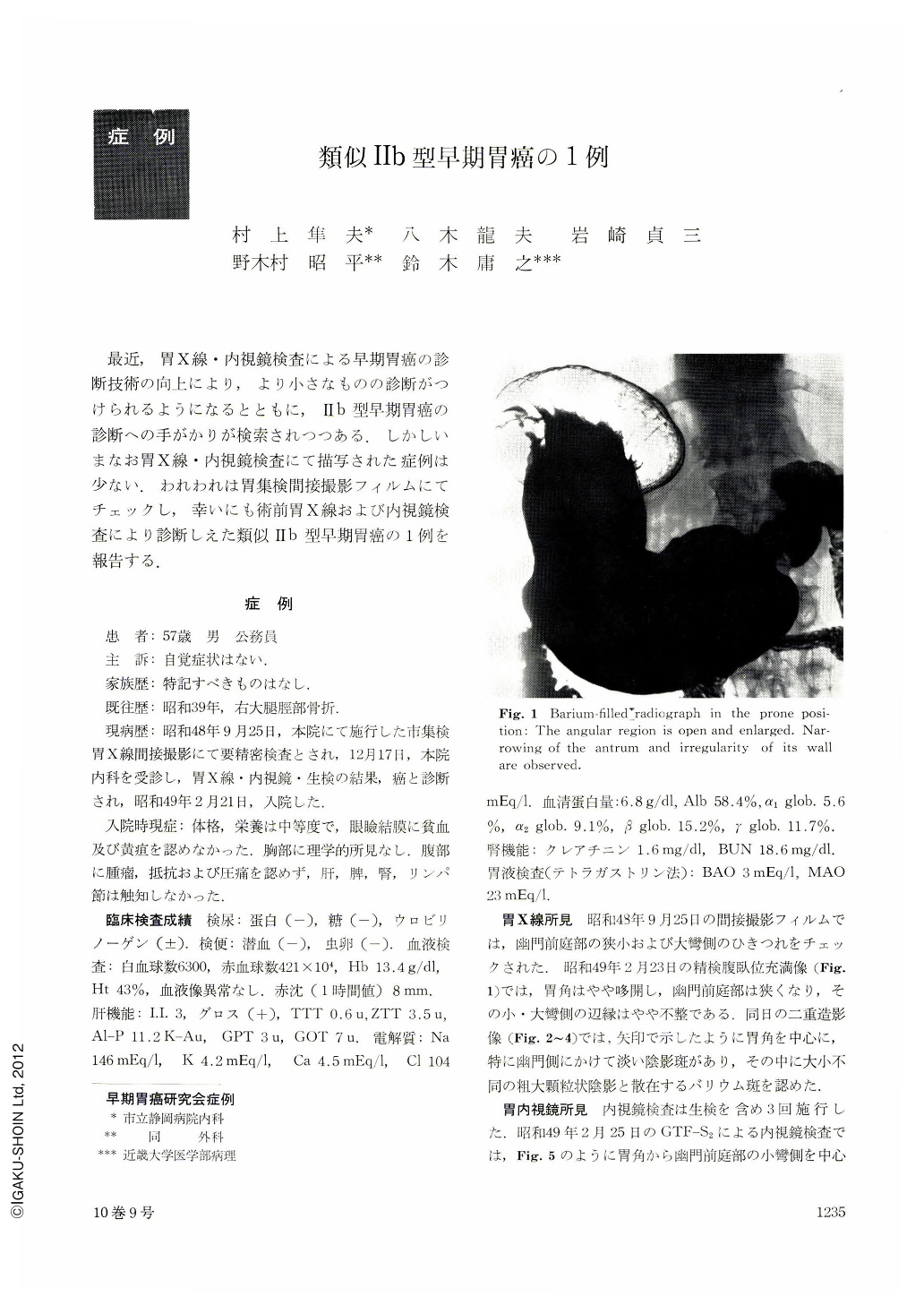
最近,胃X線・内視鏡検査による早期胃癌の診断技術の向上により,より小さなものの診断がつけられるようになるとともに,Ⅱb型早期胃癌の診断への手がかりが検索されつつある.しかしいまなお胃X線・内視鏡検査にて描写された症例は少ない.われわれは胃集検間接撮影フィルムにてチェックし,幸いにも術前胃X線および内視鏡検査により診断しえた類似Ⅱb型早期胃癌の1例を報告する.
At a gastric mass screening a 57-year-old public employe was found to harbor an abnormality in the pyloric antrum. He was then hospitalized as roentgenography, endoscopy and biopsy had led to a diagnosis of gastric cancer. Admission X-ray in barium-filled picture revealed slight widening of the angle, narrowing of the pyloric antrum and likewise slight irregular contours of both curvatures in this region. Double contrast picture showed pale flecks of shadow defect around the angle, especially in the pyloric side. Rough granular shadows of unequal size were seen in the center along with scattered barium flecks. Endoscopy revealed mottles of discolored mucosa around the angle and pyloric antrum. Reddened spots were also seen here and there, but the discolored area was ill-defined. Endoscopy prior to admission had also shown bleeding spots on the lesser curvature of the angle along with discolored mucosa there. Biopsy of these parts had resulted in the diagnosis of adenocarcinoma tubulare mucocellulare. In the resected specimen was seen a faded area centering on the lesser curvature. Fading was apparent as compared with the color of the mucosa in the mid-body. Reddened and rough mucosal picture was also seen in the pyloric side. However, the oral margins of cancerous area were ill-defined. This area, measuring 65×80 mm, extended from the angle to the pyloric antrum, surrounding the lesser curvature. It was partially exposed on the mucosal surface. While the exposed part was chiefly of tubular adenocarcinoma, the unexposed area consisted mostly of mucocellular carcinoma, CAT Ⅲ SAT, 3, with its depth m. The mucosa of non-cancerous areas showed a high degree of atrophy and intestinal metaplasia was marked in the epithelia of the gastricpits. Cut surface of the specimens did not reveal any difference in height between the area of cancer and that of normal mucosa.
Copyright © 1975, Igaku-Shoin Ltd. All rights reserved.