Japanese
English
- 有料閲覧
- Abstract 文献概要
- 1ページ目 Look Inside
症例
患 者:70歳女
主 訴:下肢の浮腫
家族歴:特記すべきものなし
既往歴:69歳で交通事故
現病歴:1977年4月頃より,両下肢の浮腫を訴え本院へ来院,低蛋白血症を指摘され,精査加療のため同年6月27日入院した.
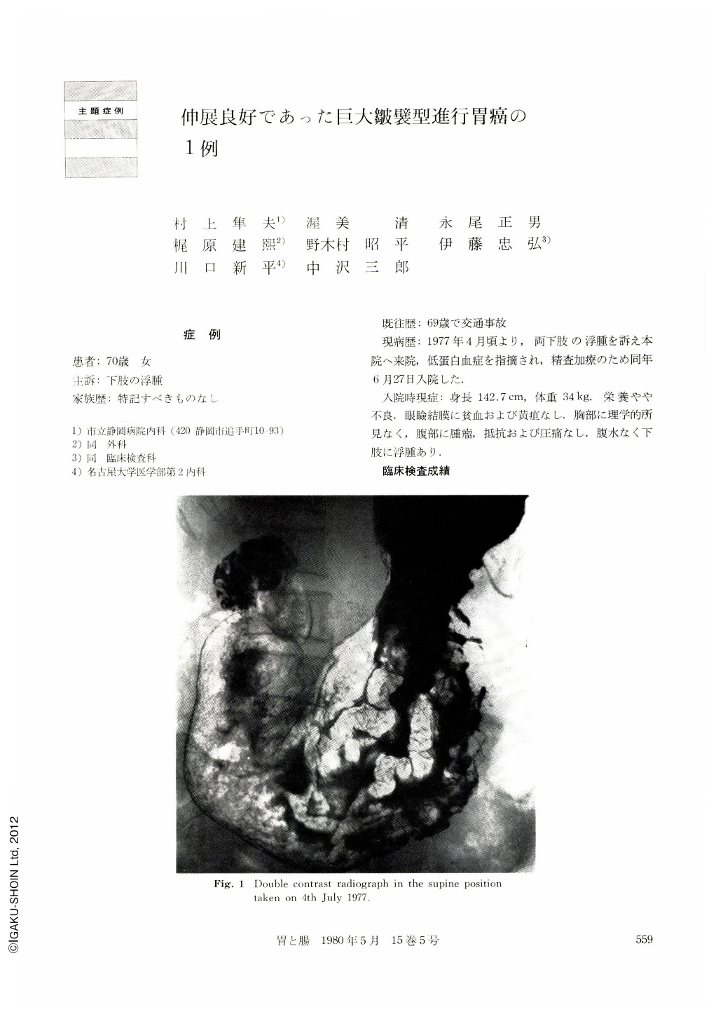
A 70 year-old woman visited our hospital with a chief complaint of edema in the legs. At admission edema was noticed in the legs and laboratory examination showed a total protein Of 4.6/dl. 1-“IPVP test was 5.4 per cent. In the upright barium-filled stomach by examination done on June 4, 1977, the lesser curvature showed irregular contour with saw-tooth changes on the greater curvature. Double contrast study showed irregular elevations over the areas of the body and the angle. In the greater curvature side were seen convolutional mucosal folds. The areae in the antrum were slightly rough but they were not exactly abnormal. Endoscopic examination on June 6 showed from the fornix down to the angle region strikingly thick and tortuous mucosal folds that were most prominent in the side of the greater curvature. The gastric wall was pliable enough and there was any change suggesting malignancy. Apparently the antrurn was normal. Fivepoint biopsy was negative for cancer. Under a diagnosis of probable Menetriez-'s disease with due allowance for diffuse infiltrative cancer we recommended strongly surgical resection. The patient left the hospital of her own will on ]uly 11. She was lost to the follow-up. In April of the following year she came to us bitterly complaining of anorexia and sensation of fulness in the abdomen. On April 25 the double contrast x-ray study of the stomach showed clearly giant mucosal folds. The gastric wall distended well. But the patient died of carcinornatous peritonitis on May 20 of the same year.
The specimen of the stomach at autopsy showed from the regions of the fornix down to the angle abnormal mucosa] folds and gyri-like findings. Moreover, on the lesser curvature of the antrum adjacent to the above area was seen an irregular-shaped excavation, measuring 19 by 16 mm, accompanied with rnucosal convergency. The nearest part of the several folds showed their abrupt cessation and wormeaten picture. Histologically, the depressed part revealed thick infiltration of signet ring cells extending from the atrophied mucosal surface layer even into the serosa. The depression was accompanied by ulcer. The entire gastric mucosa was hypertrophied. The epithelia of the gastric pits showed striking hypertrophy. The surface layer of the mucosa was hardly affected by cancer infiltration. Fibrosis of the submucosal layer, edematous and showing much dead space, was slight. Diffuse infiltration of signet ring cells were seen in the muscle layer, which was interrupted here and there by marked fibrosis. Histologic type was signet ring cell carcinoma. Its invasion partly reached beyond the serosa. The chief reason of well-kept distensibility of the wall was considered to lie in the relatively slight degree of fibrotic changes in the submucosal layer.

Copyright © 1980, Igaku-Shoin Ltd. All rights reserved.


