Japanese
English
- 有料閲覧
- Abstract 文献概要
- 1ページ目 Look Inside
- サイト内被引用 Cited by
胃X線診断や内視鏡診断技術の向上により肉眼的分類が不可能な早期微小胃癌まで術前に発見されるようになってきた.今回,われわれは,胃X線診断でその存在を疑い,内視鏡および生検で術前に診断し得た3×3mmという微小Ⅱc類似Ⅱb型早期胃癌を経験したので報告する.
A minute early gastric cancer of Ⅱb type, measuring only 3 by 3 mm, was preoperatively diagnosed by endoscopy and aiming biopsy. The lesion had been first found in the course of roentgenological examination.
The patient, a 45-year-old woman with a chief complaint of epigastric discomfort, was pointed out to have an abnormality in the greater curvature of the antrum by a roentgenological examination. She was referred to the author's hospital.
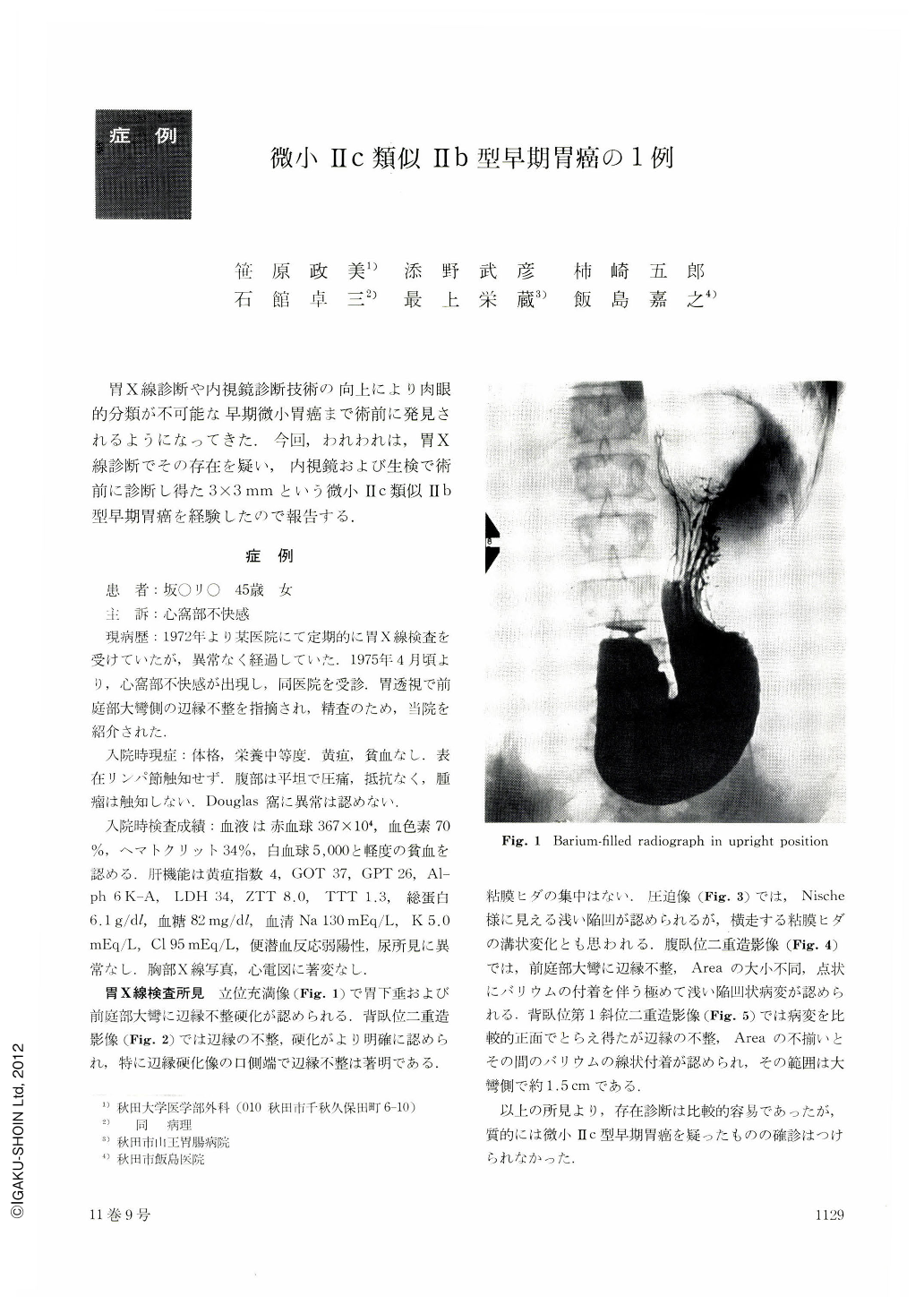
On admission, barium-filled radiograph in upright position showed irregularity of the greater curvature of the antrum. Double contrast radiograph in supine position showed irregularity and regidity of its wall more clearly, especially in the oral side. Double contrast radiograph in prone position demonstrated areae gastricae of unequal size and faint barium flecks suggesting a small depressed area. In double contrast radiograph in supine first oblique position, irregular areas and linear barium flecks in between were noticed.
Endoscopical study revealed a protrusion slightly engorged, measuring about 5 mm in diameter, on the greater curvature of the antrum. Of 6 pieces of biopsy speicimens, 2 showed Group Ⅳ and operation was performed.
Gross finding of the resected stomach in fresh and half-fixed specimens showed an erosive lesion on the greater curvature of the antrum.
Histologically, the lesion was papillary adenocarcinoma, measuring only 3 by 3 mm, localized within the mucosal layer.

Copyright © 1976, Igaku-Shoin Ltd. All rights reserved.


