Japanese
English
- 有料閲覧
- Abstract 文献概要
- 1ページ目 Look Inside
早期胃癌に対する認識が高まるにっれⅡc,Ⅱa等の陥凹型や隆起型の早期癌はかなり小さなものも術前に診断しうるようになったが,表面平坦型のⅡb病変については,中山ら1),白壁ら2)の集計にみるごとくその大部分は,癌や潰瘍,異型上皮その他の合併病変によって手術し,組織学的検索の結果偶然に発見されたものが多い.我々は,X線内視鏡,生検により術前にⅡbと診断しえた広範囲な単独Ⅱb病変の一例を経験したので報告する.
A 67-year-old woman came to the hospital with complaints of weight loss and epigastric fullness of a few months' duration.
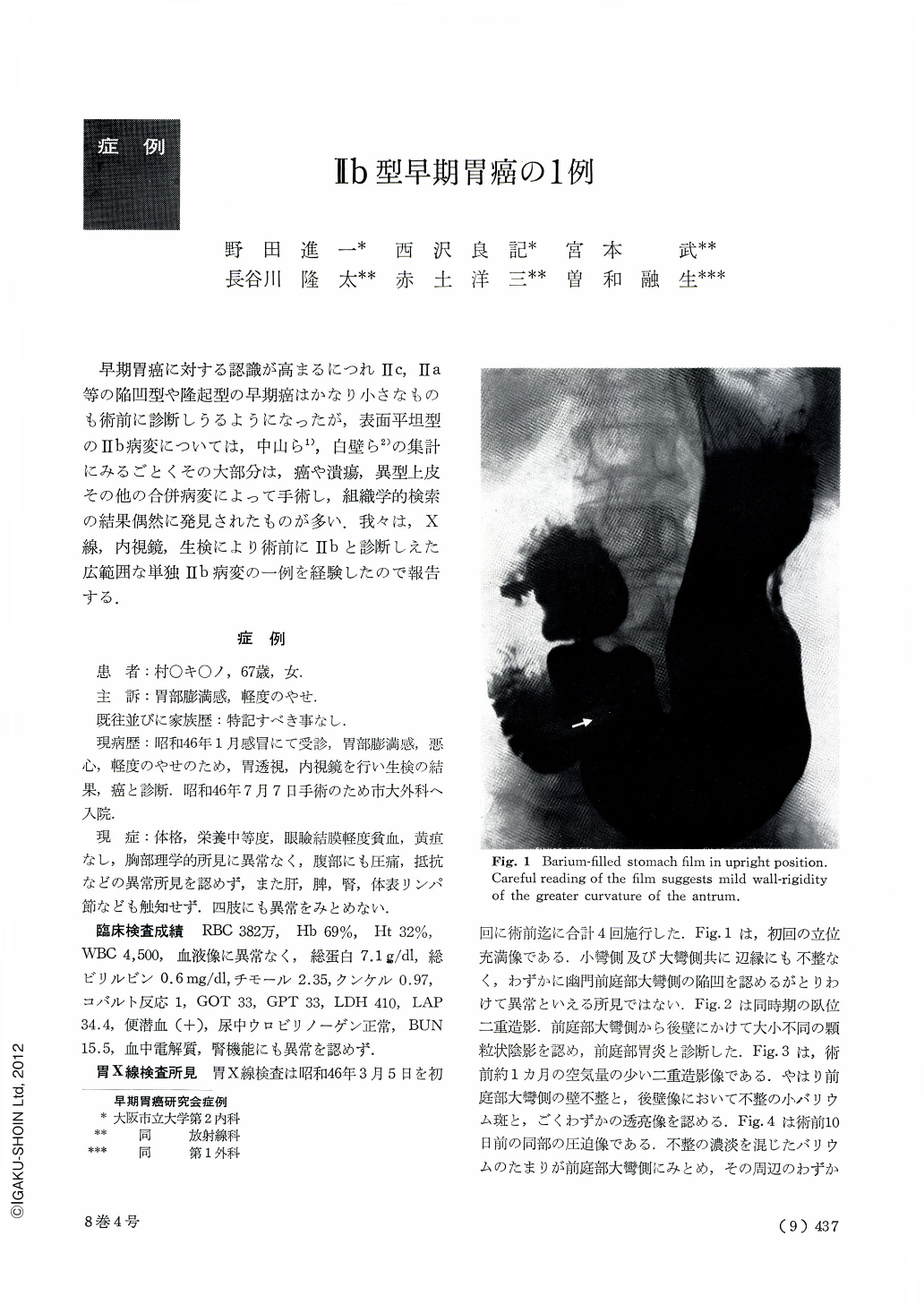
The films of the first upper GI series revealed that the greater curvature and adjacent posterior wall of the antrum showed barium coating slightly different from the other areas and this area was characterized by small and large granular appearance of the mucosa.
Reddish fleck was seen at the greater cuvature of the antrum on the first gastrocamera examination and the possibility of Ⅱc type carcinoma could not be excluded. The gastrofiberscopic examination done one month later showed the similar reddish fleck and another irregular, elevated area at the posterior wall of the antrum which were suspected of being Ⅱc type cacinoma. Biopsy was done on these places. Signet ring celles were found in the five out of six biopsied specimens.
Preoperative upper GI series showed poorly defined accumulation of barium at the antrum on compression study and indistinct barium fleck on double contrast study, but the areas of carcinomatous invasion usually seen in Ⅱc type carcinoma could not be traced.
Preoperative fiberscopic examination with small amount of air showed irregular, discolored, coarse gastric mucosa on the posterior wall of the antrum. The coarseness of the mucosa disappeared by stretch-ing its surface with large amount of air. Only discoloration remained.
These findings suggested Ⅱb type carcinoma. In the fresh as well as fixed gross specimens of the resected stomach, there was an elevated area with rough irregular surface seen on the antrum in the greater curvature which was probably made by thickening and fusion of the “gastric areas”.
The surface of the mucosa around the elevated area was without brightness, partly coarse and its “gastric areas” were poorly defined, but the area of carcinomatous invasion could not be traced after all.
Adenocarcinoma mucocellulare was histopathological diagnosis, and the extent of carcinomatous invasion into the deep layers was “m”.
The thickness of the mucosa invaded by cancer was the same as that of normal mucosa and there were a few small erosions of the mucosa. The lesion was band-form Ⅱb type with the dimensions of 2.7×9.0cm, extending widely from the greater curvature to the anterior and posterior wall of the antrum. This is a case of Ⅱb subtype early gastric cancer in which mucosal change could be demonstrated radiologically and endoscopically and diagnosis of cancer could be made by biopsy. Cases with widespread Ⅱb type carcinoma like this case have been rarely reported in the literature and this might be a case valuable in considering the pathogenesis of gastric cancer.

Copyright © 1973, Igaku-Shoin Ltd. All rights reserved.


