Japanese
English
- 有料閲覧
- Abstract 文献概要
- 1ページ目 Look Inside
筆者らは,術前胃X線,内視鏡により早期胃癌を疑い,結局潰瘍を合併した胃角副膵であった1例を報告する.
症例
患 者:61歳 男子 教員
主 訴:空腹時心窩部重圧感および食後胃部膨満感.
家族歴:特記すべきものなし.
既往歴:特記すべきものなし.
現病歴:数年前より,時々空腹時に心窩部重圧感を訴えていた.昭和46年5月初旬頃よりその程度が増強し,食後胃部膨満感もきたすようになったので某医を受診し胃X線にて,幽門狭窄を指摘され,昭和46年6月21日精査のため,本院へ入院した.
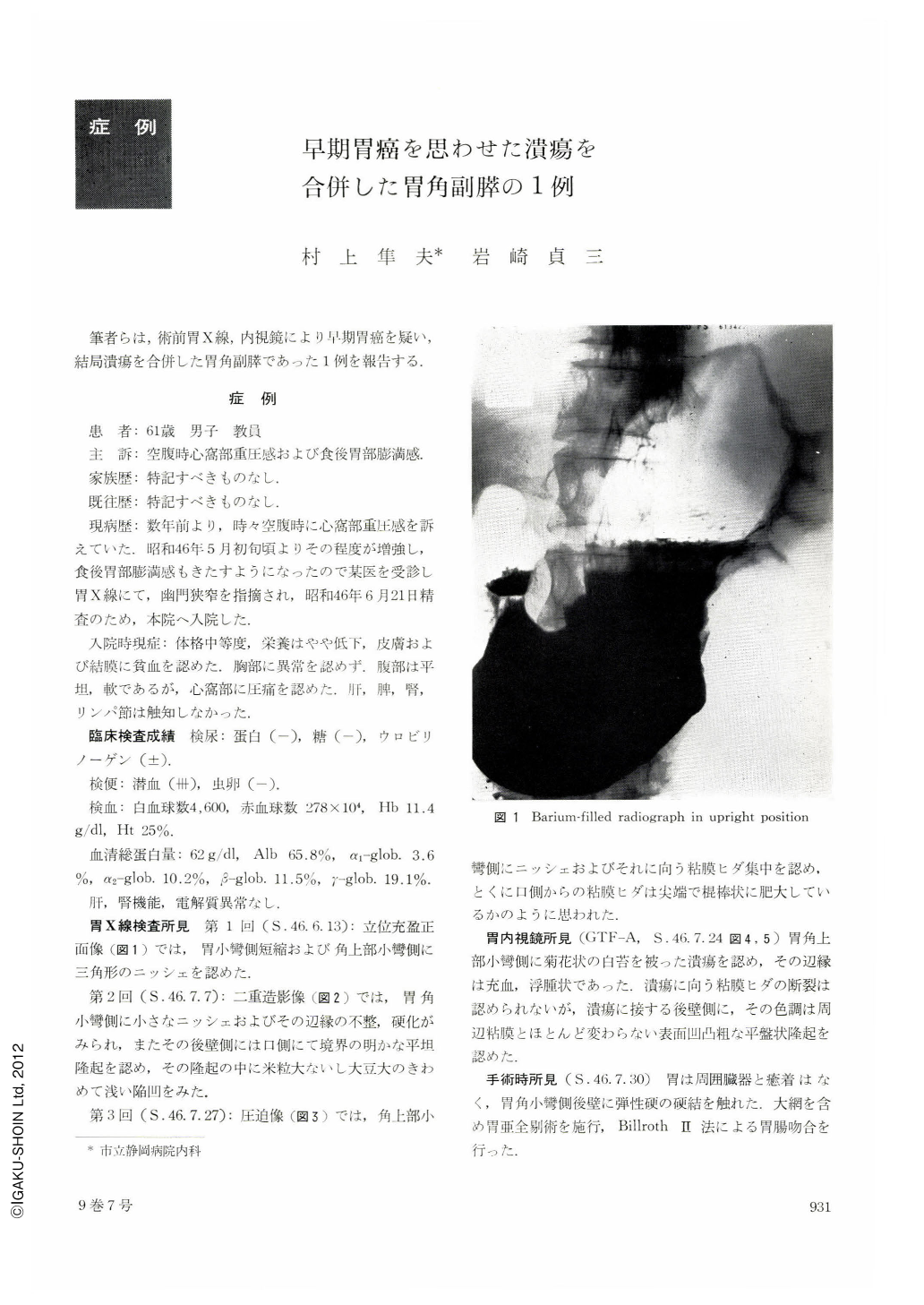
A 61-year-old man was admitted to hospital complaining of epigastric fullness from which he suffered since several years before. On admission he was emaciated with tenderness in the epigastrium. Laboratory examination showed anemia and the feces was positive for occult blood. Double contrast method in x-ray revealed a niche, irregularity and marginal rigidity at the angle of the lesser cuvature. There was a well-defined flat elevation on the oral side at the posterior wall adjacent to the niche. Endoscopic examination also showed an open and irregular ulcer with mucosal convergence running from the anterior wall at the level of the angle. The converging mucosal folds did not exhibit any clubbiness or fusion at the tips. There was seen reddish swelling around the ulcer as well as a flat elevation with pale coarse granules at the posterior wall adjacent to the ulcer.
The operation done under a provisional diagnosis of early stomach cancer macroscopically revealed an irregular ulcer, 20 × 10 mm, at the angle of the lesser curvature, accompanied with a slightly thick mucosal convergence. The flat elevation on the posterior wall adjacent to the ulcer looked normal in both consistency and color.
An aberrant pancreatic tissue was histologically seen located under the flat elevation and a part of the ulcer, extending to the serosa through the submucosal layer. It simulated a normal pancreatic tissue. Orifices of some pancreatic ducts were open above the surface of the ulcer. It is interesting that pancreatic juice supposedly induced the ulcer by its digestive process.

Copyright © 1974, Igaku-Shoin Ltd. All rights reserved.


