Japanese
English
- 有料閲覧
PPV購入案内
1,320円 (1,200円+税10%) こちらは、239ページから244ページの文献です。購入には医書.jp本会員登録が必要です。
会員登録完了後に改めて上記「購入サイトへ」を選択してください。
または「購入サイトへ」選択後に「購入ログイン」、「新規会員登録」の順に進んでください。
会員登録について詳しくはをご確認ください。
- Abstract 文献概要
- 1ページ目 Look Inside
胃X線診断能の向上とともに,近年の胃内視鏡診断能の向上には著しいものがあり,胃疾患に対する形態学的な診断学はほぼ完成の域に達したといっても過言ではない.なかでも胃内視鏡検査の補助的診断法として,色素撒布法が用いられるようになってからは,胃粘膜面の微妙な色調差や微細凹凸変化を的確に観察できるようになり,微小早期胃癌の内視鏡診断も決して困難ではなくなった.最近この内視鏡的色素撒布法を用いて,術前に診断可能であった広範なⅡb型早期胃癌症例を経験したので報告し,あわせて色素撒布法の有用性について検討してみた.
Recently we have come across an extensive Ⅱb type early gastric cancer that was diagnosed preoperatively by means of endoscopic dye scattering method. Here is its report together with some comments on the efficacy of this method.
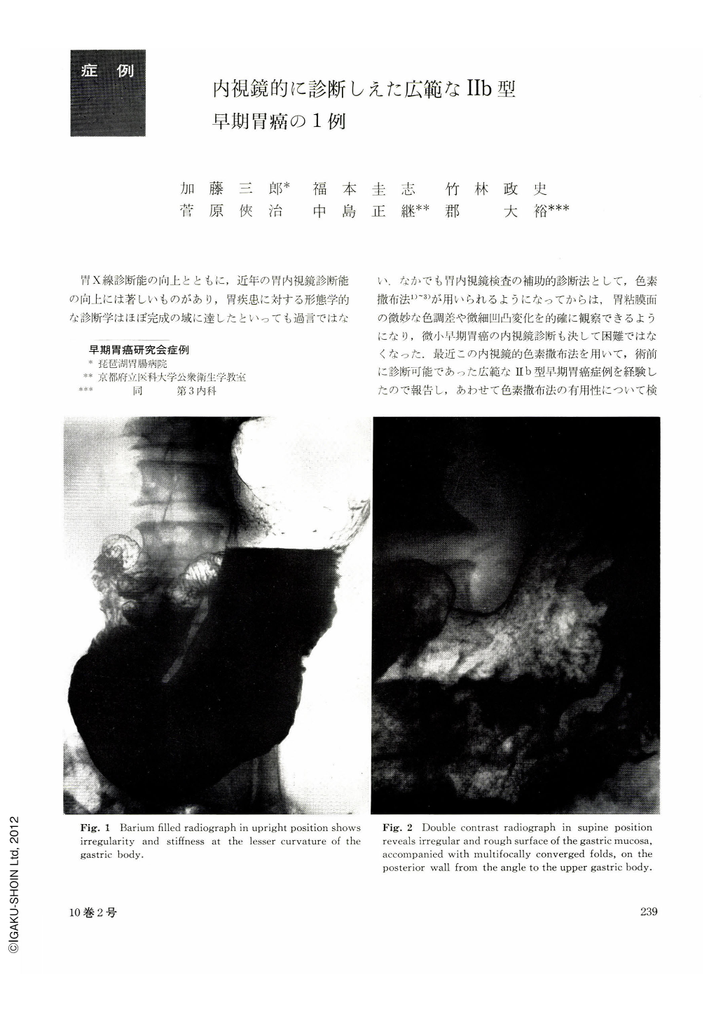
The patient, a 48-year-old man, visited our hospital for thorough workup because of deformity of the duodenal bulb detected at a gastric mass screening. Roentgenographic examination of the stomach revealed in the upright barium-filled picture slight irregularity and rigidity of the gastric contour on the lesser curvature of the body. In double contrast picture the same contour looked fluffy and twofold. On the posterior wall of the body were seen extensive areae gastricae of varying size with partial obliteration. These findings led us to suspect extensive flat early gastric cancer (Ⅱb) and endoscopy was subsequently performed. There were reddened spots here and there with adhesion of thin white coats on a wide area from the upper body down to the gastric angle with the lesser curvature as its center. As histological diagnosis of undifferentiated carcinoma was obtained by biopsy, endoscopic dye scattering method was done in order to determine the extent of malignancy. Scattering with indigocarmine emphasized the mucosal reddening, clarifying minute unevenness of the mucosal surface, while scattering with methylene blue showed deeper staining of the rough areas which then looked depressed as compared with the surrounding mucosa. The resected specimen showed that the surface of the depressed area looked flat and smooth, dotted with sporadical reddenings and rough granules. In the fixed specimen the diseased area looked ironed-out with disappearance of the areae gastricae, easy to differentiate from the surrounding normal mucosa. However, histologically the area of cancer, which proved to be signet ring cell carcinoma, was narrower than that of the preoperative diagnosis. It was 60 by 50 mm in dimensions, partially exposed over the mucosal surface but mostly localized within the middle layer of the mucosa.
In the diagnosis of Ⅱb type early cancer, especially of cancer lesions exposed over the mucosal surface, dye scattering method is effective in making out difference of mucosal tone such as reddening or minute unevenness of the surface. At the same time, intramucosal carcinoma, especially when it is covered with epithels of intestinal metaplasia, methylene blue scattering stains the diseased area in the same manner as the normal mucosa. This should always be borne in mind in the practice of dye scattering method.
Copyright © 1975, Igaku-Shoin Ltd. All rights reserved.