Japanese
English
- 有料閲覧
- Abstract 文献概要
- 1ページ目 Look Inside
十二指腸に原発する良性腫瘍はきわめて稀とされており,中村ら1)によると,欧米では十二指腸の良性腫瘍280例中腺腫が113例(40%)と最も多く,Brunner腺腫が45例(16%)とこれに次いでおり,本邦では逆にBrunner腺腫が56例中25例(45%)と最も多く,腺腫は10例(18%)となっている.以前にわれわれが検討した本邦例の集計でも2),良性十二指腸腫瘍38例中Brunner腺腫が16例(42%)と最も多かった.しかし,いずれもX線診断ないしは剖検時の観察によって発見された症例ばかりであり,最近十二指腸への内視鏡検査法が確立され,広く応用されるようになってみると,微細な病変まで含めると,十二指腸の隆起性病変は従来報告されているほど頻度の低いものではないことがわかってきた.
今回,われわれは最近経験したBrunner腺腫2例について報告し,併せて若干の文献的考察を試みた.
In this paper are reported two cases of brunnerioma of the duodenum as its tumors of benign nature are encountered only very infrequently. We have also collected the literature hitherto published in Japan, with some comments on their incidence, site of origin signs and symptoms caused by them and their pertinent diagnosis.
Case 1: a 25-year-old man.
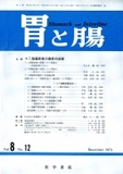
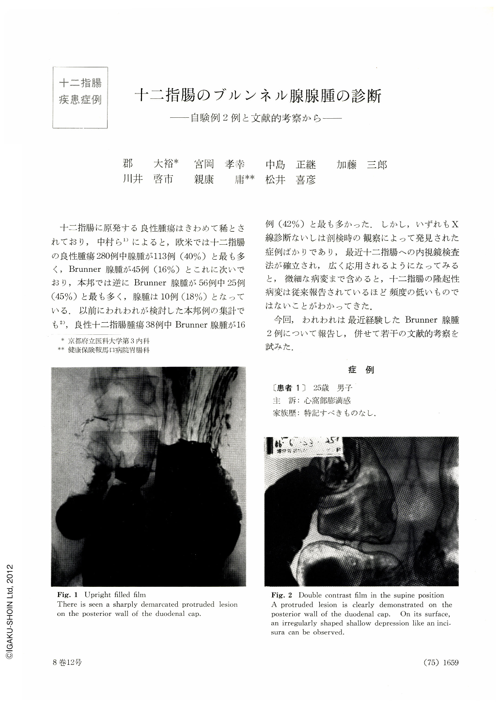
He visited our hospital with a chief complaint of sensation of fullness in the epigastrium. Fluoroscopy of the upper digestive tract revealed a well-defined shadow defect with smooth surface the size of a small finger tip located on the posterior wall of the duodenal bulb. Endoscopy showed in the same site a small tumor also the size of a small finger tip with a short stalk. The surface of the neoplasm as a whole was of normal color and smooth, but the central part way reddened with shallow depression. Boring biopsy of the center revealed massive hyperplasia of Brunner's glands beneath atrophied duodenal mucosa. Brunnerioma, suspected by biopsy, was confirmed as such after its surgical extirpation. It measured 13 by 10 mm.
Case 2: a 70-year-old man.
His subjective symptoms were not remarkable. At a gastric mass screening he was found to harbor polyposis of the duodenal bulb and was admitted to the hospital for thorough check-up. Detailed x-ray revealed on the anterior wall side of the bulb a sharply demarcated shadow defect with the shape of a green caterpillar along with several similar other shadow defects, round or oval, in the same location. Endoscopy disclosed over the whole circumference of the duodenal wall several well-defined reddened protrusions. The largest one had the shape of a green caterpillar with small erosions covered with white coat over it. As in Case 1. strong suspicion of brunnerioma was entertained by biopsy, and gastrectomy was performed. Histologically, there were 5 protrusions including the largest one, which measured 25 by 7 mm in its largest diameters. All protrusions consisted of hyperplasia of the Brunner's glands chiefly distributed in the submucosal layer. The lamina propriae was raised in the manner of a fountain or a box.
Our results of investigation regarding roentgenologic and endoscopic diagnosis of brunnerioma partly based on the present cases suggest that some of brunneriomas are hard to discriminate from other non-epithelial tumors, but yet when brunnerioma reaches to a certain size its differentiation is not so difficult.
Copyright © 1973, Igaku-Shoin Ltd. All rights reserved.