Japanese
English
- 有料閲覧
- Abstract 文献概要
- 1ページ目 Look Inside
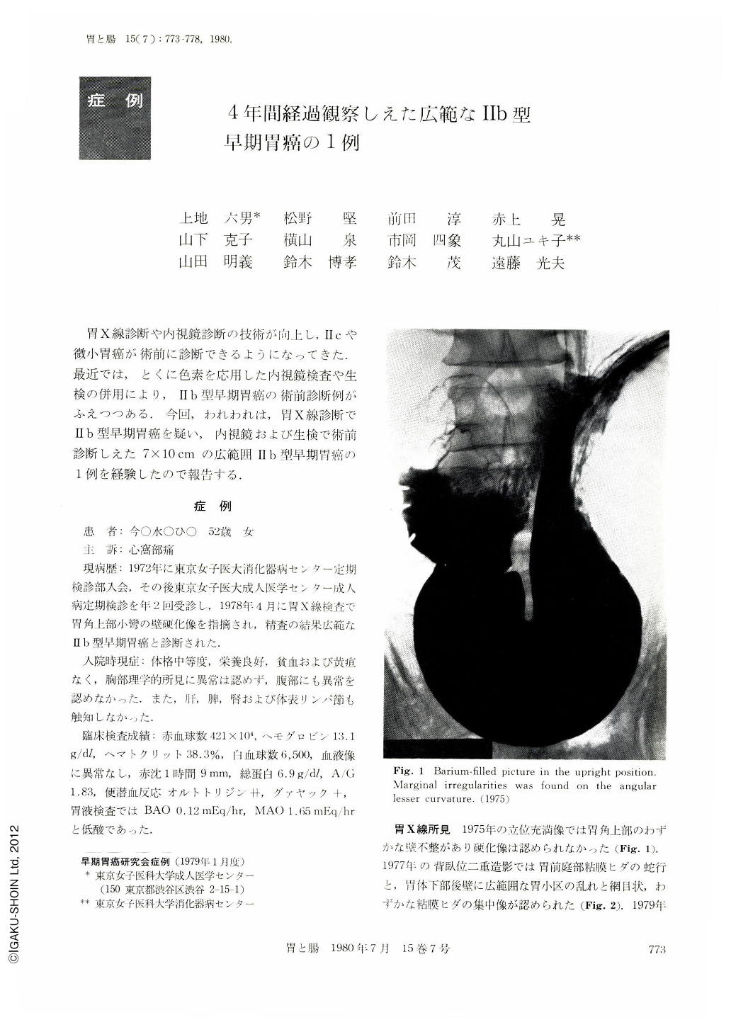
胃X線診断や内視鏡診断の技術が向上し,Ⅱcや微小胃癌が術前に診断できるようになってきた.最近では,とくに色素を応用した内視鏡検査や生検の併用により,Ⅱb型早期胃癌が術前診断例がふえつつある.今回,われわれは,胃X線診断でⅡb型早期胃癌を疑い,内視鏡および生検で術前診断しえた7×10cmの広範囲Ⅱb型早期胃癌の1例を経験したので報告する.
症例
患 者:今○水○ひ○ 52歳 女
主 訴:心窩部痛
現病歴:1972年に東京女子医大消化器病センタ一定期検診部入会,その後東京女子医大成人医学センター成人病定期検診を年2回受診し,1978年4月に胃X線検査で胃角上部小彎の壁硬化像を指摘され,精査の結果広範なⅡb型早期胃癌と診断された.
The patient is 52 year-old woman. X-ray examination of the stomach showed irregular areae gastricae on the anterior and posterior walls, centering on the lesser curvature above the angle. Endoscopy also showed on the same place white discolored flecks. From these findings we made a diagnosis of extensive Ⅱb type early cancer, centering on the part above the angle. Macroscopically the resected stomach showed whitish flat lesion, well-defined and measuring 7 by 10 cm, with the lesser curvature above the angle in its center. Histologically were seen in a part of the lesser curvature above the angle well differentiated and moderately differentiated adenocarcinoma lesions. In the other parts including the outer margins of the whole lesion was seen poorly differentiated adenocarcinoma. No metastasis was recognized.
The patient was followed up for four years. At the initial x-ray examination of the stomach no abnormality was found and two years later gastric lesion was found ultimately by both x-ray and endoscopy. Especially reddened flecks as revealed by endoscopy clinched the diagnosis. When we put findings of the resected specimen and its histologic changes all together, the lesion was surely of typical Ⅱb. The site of engorged flecks found two years later since the initial examination corresponded histologically to that of well differentiated adenocarcinoma. The superficial layer was replaced by intestinal metaplasia, showing atypical changes. We assumed that such histologic findings were recognized here because there had had been a Ⅱc-like lesion here and it had healed completely. In other parts were found poorly differentiated carcinoma and signet-ring-cell carcinoma. We also assumed that the extensive lesion had spread gradually and taking a long time in the superficial layer with the Ⅱc lesion on the lesser curvature above the angle as the starting point of carcinomatous changes. The present case made us aware once again of the fact that how important are the engorged and discolored flecks as revealed by endoscopy in the diagnosis of Ⅱb lesion.

Copyright © 1980, Igaku-Shoin Ltd. All rights reserved.


