Japanese
English
- 有料閲覧
PPV購入案内
1,320円 (1,200円+税10%) こちらは、367ページから369ページの文献です。購入には医書.jp本会員登録が必要です。
会員登録完了後に改めて上記「購入サイトへ」を選択してください。
または「購入サイトへ」選択後に「購入ログイン」、「新規会員登録」の順に進んでください。
会員登録について詳しくはをご確認ください。
- Abstract 文献概要
- 1ページ目 Look Inside
患 者:66歳 女性
主 訴:悪心および嘔吐.
既往歴:便中に回虫卵を指摘されたことあり.
家族歴:特記すべきことなし.
現病歴:約2週間前より軽度の心窩部痛と共に悪心および嘔吐を認め,昭和47年5月6日琵琶湖胃腸病院を受診.
現 症:心窩部に圧痛を認める以外に著変なし.
一般検査所見:便に回虫卵を認める以外に血液検査,肝機能検査,その他諸検査に異常なし.
Case: a 66-year-old woman.
Chief complaints: nausea and vomiting.
Past history: At one time the feces was positive for ascaris ova.
Familial history: non-contributory.
Present history: Since about two weeks before she had slight epigastrial pain along with nausea and vomiting, and on May 6, 1972, she visited the Biwako Gastroenterological Hospital.
Status praesens: No remarkable changes except tenderness in the epigastrial region.
General examinations: Except ascaris ova in the feces various examinations including the blood, liver function tests were within normal limits.
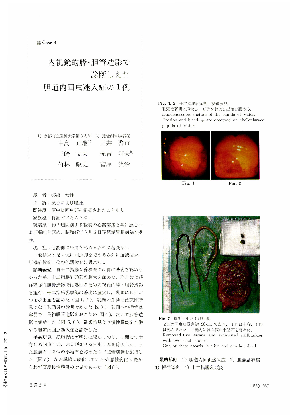
Course of the diagnosis: Upper gastrointestinal x-ray series showed no abnormality in the stomach, and swollen papillary region in the duodenum. As both peroral and intravenous cholecystography disclosed nothing unusual, endoscopic pancreatocholangiography was attempted. The papillary region of the duodenum was markedly swollen, with erosions and bleeding on the papilla of Vater (Fig. 1 and 2).
Biopsy of the papilla revealed no malignant changes. Diagnosis then was papillitis (Fig. 3). Cannulation into the papilla was not diffcult, and first the pancreatic duct was opacified (Fig. 4), followed by positive cholangiography (Fig. 5 and 6). Findings of the visualized ducts led us to a diagnosis of biliary ascariasis associated with chronic pancreatitis.
Findings at operation: Dilatation of the common bile duct was pronounced. By incision an alive ascaris and another dead one were taken out of the common bile dudt. Incidentally, two small stones were seen in the gallbladder, so that cholecystectomy was performed (Fig. 7). The pancreas was cirrhotic, but no malignant changes were observed. Findings obtained were thus indicative of advanced chronic pancreatitis.
Final diagnosis: 1) bile duct ascariasis.2) cholelithiasis. 3) chronic pancreatitis. 4) duodenal papillitis.
Copyright © 1973, Igaku-Shoin Ltd. All rights reserved.