Japanese
English
- 有料閲覧
- Abstract 文献概要
- 1ページ目 Look Inside
幽門近旁部は粘膜の萎縮および腸上皮化性を強く来し,さらにビランや潰瘍などの修復に伴う変形を生じやすく,その上,胃の旺盛な蠕動運動の影響をうける部位である.それにX線検査では,十二指腸とか脊柱との重なりのために鮮明な描出が困難で,また内視鏡検査でも従来は変形とか遠景のために目的部位の観察が充分でなかったり,生検も不正確に終ることが多かった.したがって悪性病変を疑いながらも確診を得るに至らないままに胃切除術を施行したが,その後の組織学的検索にて良性であったものや,悪性を強く疑いながらも確診できず,そのまま経過観察のやむなきにいたった症例も経験してきた.
しかし,最近の側視式十二指腸ファイバースコープ(JF-B),またGIF-Dを中心とした直視式のPanendoscopeの開発により,幽門およびその近傍部を直視下に正面像をとらえ,詳細な観察と正確な生検により,幽門近旁部病変の診断を正確に,とりわけ微小胃癌の術前診断をも可能にした.
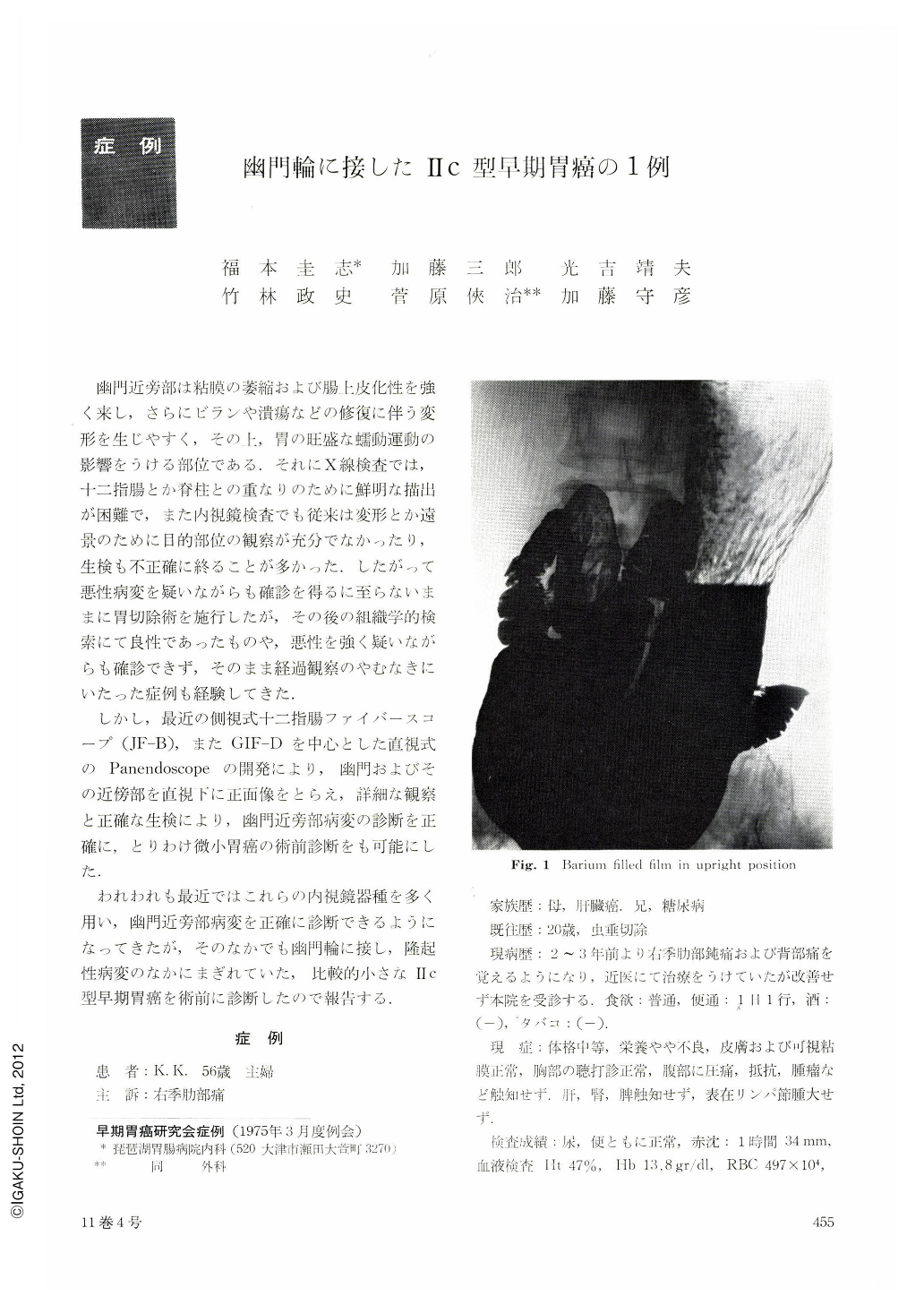
A woman aged 56 came to our hospital with a chief complaint of dull pain in the right hypochondrium. X-rav revealed two protruding lesions the size of a pea and red bean, respectively, in the neighborhood of the pylorus and another couple of elevations in the duodenum.
Endoscopy with GTF-SII showed two protrusions each in the lesser curvature side and on the posterior wall. The intervening area on the posterior wall was hemorrhagic. The bleeding spot was recognized by JF-BII as a depression. A IIc lesion was then suspected. Also were seen in the duodenal bulb two smooth-surfaced protrusions of varying size. Minute observation of the surface of the two protrusions in the pylorus suggested their benign nature. On the other hand, when dye scattering method was coemployed, the extent of depression in the pylorus and rough granular changes around it were seen to better advantage and malignancy was suspected. Of 11 pieces of biopsy specimens taken from all these parts, well differentiated carcinoma was recognized only in the depression. Resected specimen showed, corresponding to the endoscopic observations, an irregularshaped depression accompanied with reddening on the posterior wall side in the neighborhood of the pyloric ring along with two elevations there.
Histologic study showed differentiated tubular carcinoma, 25×7 mm in diameters, with depth degree m, in an area corresponding to the irregular-shaped depression. It was also shown that the protrusion in the lesser curvature side was an adenomatous polyp, and rough granulas on the posterior wall represented intact mucosa left within the Ⅱc area. Protrusions in the duodenal bulb proved to be Brunnerioma.
The present case illustrates the fact that preoperative diagnosis by endoscopy is becoming more accurate. X-ray diagnosis was protruding lesions in the stomach and duodenal bulb, but a small IIc type cancer did exist in between.
Copyright © 1976, Igaku-Shoin Ltd. All rights reserved.