Japanese
English
- 有料閲覧
- Abstract 文献概要
- 1ページ目 Look Inside
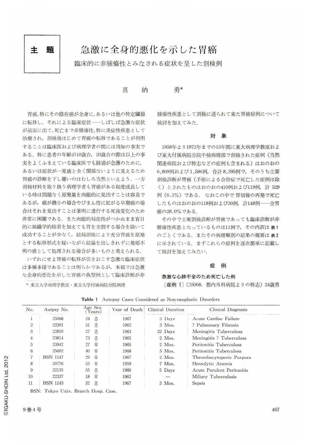
胃癌,特にその陰在癌が全身に,あるいは他の特定臓器に転移し,それによる臨床症状──しばしば急激な症状が前面に出て,死亡まで非腫瘍性,特に炎症性疾患として治療され,剖検後はじめて胃癌の転移であることが判明することは臨床医および病理学者の間には周知の事実である.特に患者の年齢が10歳台,20歳台の際は以上の事実をよくふまえている臨床医でも経過が急激のために,あるいは症状が一見癌と全く関係ないように見えるため胃癌の診断を下し難いのはむしろ当然といえよう.一方剖検材料を取り扱う病理学者も胃癌がある程度成長している時は問題なく原発巣を肉眼的に見出すことは容易であるが,癌が微小の場合やびまん性に拡がる早期癌の場合はそれを見出すことは著明に進行する死後変化のため非常に困難である.また肉眼的局在性がつかぬまま盲目的に組織学的検索を加えても胃を全割する場合を除いて成功することが少なく,結局剖検により充分胃癌を原発とする転移形式を疑いながら結論を出しきれずに発原不明の癌として処理される場合が多いものと考えられる.
いずれにせよ胃癌の転移が引きおこす急激な臨床症状は多種多様であることは明らかであるが,本稿では急激な全身的悪化を示した胃癌の典型例として臨床診断が非腫瘍性疾患として剖検に送られて来た胃癌症例について検討を加えてみた.
Eleven autopsy cases of gastric carcinoma, clinically diagnosed as non-neoplastic disorders, as representative cases demonstrating rapid generalized deterioration, were selected from the files of Department of Pathology, Tokyo University and Section of Pathology, Central Laboratry, Tokyo University Branch Hospital during the years 1958~1973. This comprised 0.13% of the total 8,395 autopsy cases and 2.1% of the total 529 autopsy cases for gastric cancer.
According to clinical manifestations the following classification was made.
1. Cardiopulmonary insufficiency
2. Meningeal irritation with increased intracranial pressure 2 cases
3. Symptoms of peritonitis 3 cases
4. Hematoporetic disorders 2 cases
5. Generalized infection 2 cases
Consequently 7 cases were considered as infection diseases (mostly tbc.) and 4 as non-infection disorders. Nine cases demonstrated clinical symptoms caused by cancer metastasis per se. One patient died of purulent peritonitis due to perforation of ulcerative gastric cancer, and the other succumbed to sepsis secondary to gastric cancer. The youngest age was 18 and the oldest 73 with the mean years of 37.4. The shortest clinical course was 3 days, in which case the diagnosis was acute cardiac failure, and the longest was 7 months with a mean value of approximately 2 1/2 months. There has been no case of gastric cancer clinically diagnosed as infections or hematopoietic diseases since 1969, suggesting great improvement of X-ray diagnosis and endoscopic techniques.
Pathologically all cases but one revealed cancer reaching the serosa. The exceptional case was early cancer, 2×2 cm in size, involving only the mucosa and submucosa, with extensive meningeal metastasis. Histologically 9 cases showed adenocarcinoma mucocellulare (undifferentiated or poorly differentiated adenocarcinoma). In the remaining two cases one showed tubular pattern, the other varied structures ranging from mucocellular to papillotubular adenocarcinoma. The latter was 18-year-old female with clinical diagnosis of miliary tuberculosis.
Clinical manifestations exactly corresponded to the main cancer metastasis, that is, cardiopulmonary insufficiency to diffuse lung carcinomatosis, meningeal irritation to diffuse meningeal metastasis; hematcpoietic disorders to extensive bone marrow metastasis. Nine patients, all having distant metastasis with or without peritoneal dissemination, demonstrated retroperitoneal lymph node involvement, most of which also revealed Virchow's meatastasis. In 6 of llcases there was diffuse pulmonary meatastasis in the form of vascular and/or lymphatic tumor embodism. In 3 and 5 cases there were diffuse meningeal and bone marrow meatstases, respectively. Of 5 cases with bone metastasis 4 had metastatic lung carcinomatosis at the same time. Because bone marrow metastasis was hematogenous and in metastatic pulmonary carcinomatosis both vascular and lymphatic involvements were present the author maintains the theory of the hematogenous nature of pulmonary lymphangitis carcinomatosa. In addition other recent investigations also support this theory.
As for the meningeal metastasis most past studies favoured lymphatic spread through the vertebral foramen transversarium partly because in most cases retroperitoneal lymph-nodes were extensively involved by cancer. However, since some reported cases had no retroperitoneal involvement and Virchow's nodes were mostly invaded by cancer, meningeal carcinomatosis might also be due to hematogeneous spread. The fact that histology in most cases with lung and bone metastasis showed similar features to that of cases with diffuse meningeal carcinomatosis also supports the theory of hematogenous metastasis.

Copyright © 1974, Igaku-Shoin Ltd. All rights reserved.


