Japanese
English
- 有料閲覧
- Abstract 文献概要
- 1ページ目 Look Inside
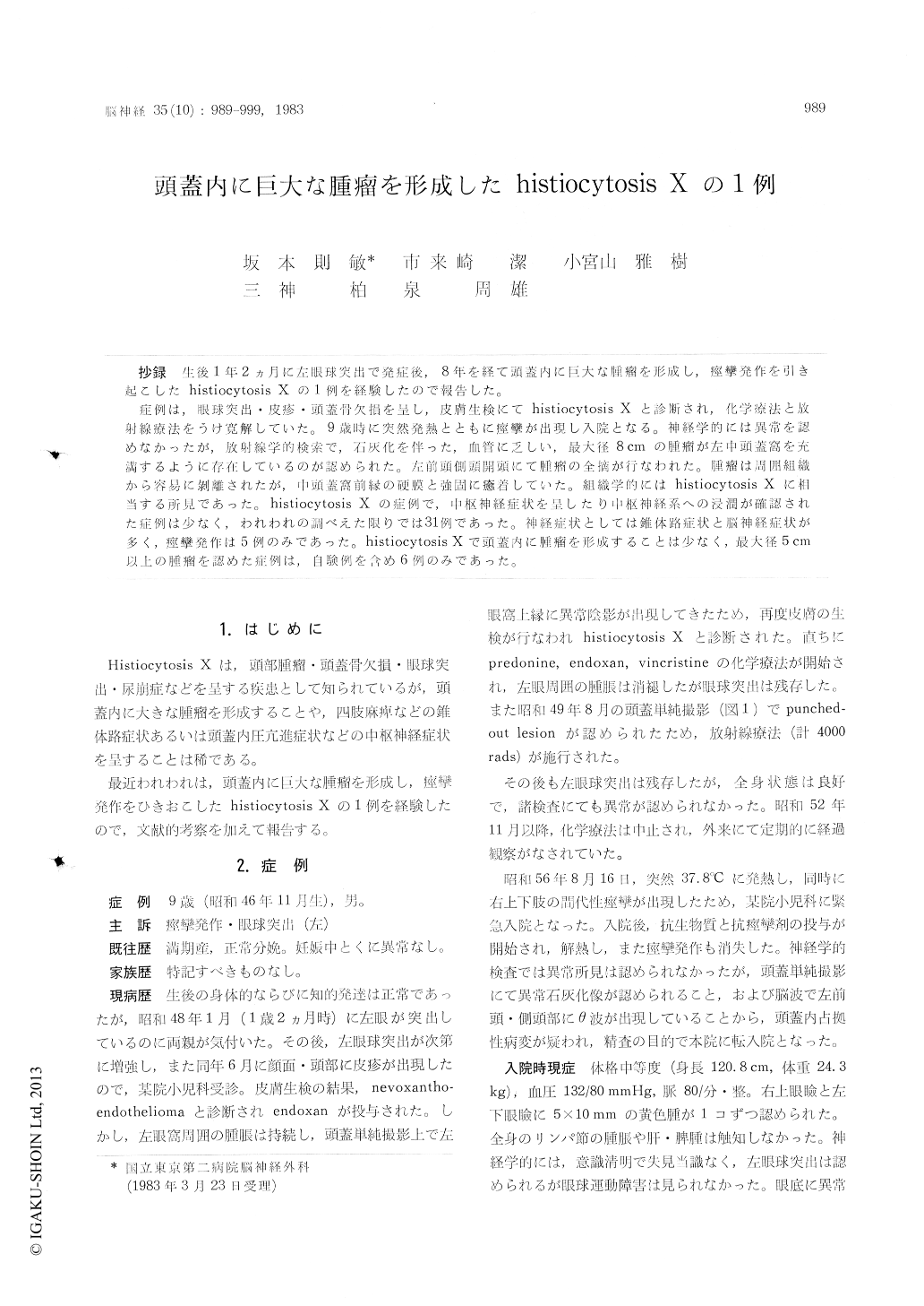
抄録 生後1年2ヵ月に左眼球突出で発症後,8年を経て頭蓋内に巨大な腫瘤を形成し,痙攣発作を引き起こしたhistiocytosis Xの1例を経験したので報告した。
症例は,眼球突出・皮疹・頭蓋骨欠損を呈し,皮膚生検にてhistiocytosis Xと診断され,化学療法と放射線療法をうけ寛解していた。9歳時に突然発熱とともに痙攣が出現し入院となる。神経学的には異常を認めなかったが,放射線学的検索で,石灰化を伴った,血管に乏しい,最大径8cmの腫瘤が左中頭蓋窩を充満するように存在しているのが認められた。左前頭側頭開頭にて腫瘤の全摘が行なわれた。腫瘤は周囲組織から容易に剥離されたが,中頭蓋窩前縁の硬膜と強固に癒着していた。組織学的にはhistiocytosis Xに相当する所見であった。histiocytosis Xの症例で,中枢神経症状を呈したり中枢神経系への浸潤が確認された症例は少なく,われわれの調べえた限りでは31例であった。神経症状としては錐体路症状と脳神経症状が多く,痙攣発作は5例のみであった。histiocytosis Xで頭蓋内に腫瘤を形成することは少なく,最大径5cm以上の腫瘤を認めた症例は,自験例を含め6例のみであった。
The authors experienced a case of histiocytosisX with a large intracranial mass resulting in a convulsive seizure. The patient showed left ex-ophthalmos and a skin rash one year and two mon-ths after birth. Histiocytosis X was diagnosed from a skin biopsy, and predonine, endoxan and vin-cristine were administered. The rash disappeared, but the exophthalmos remained. At the age of two years and nine months, punched-out lesions appeared in the skull and 4,000 rads of radiation was applied. Thereafter, the exopthalmos persisted but there was no particular problem in the course. However, a convulsive seizure with fever suddenly appeared at nine years and ten months of age and the patient was hospitalized.
At the time of admission, the general condition was good and there were no abnormalities in neu-rological tests. In neuroradiological examinations, a calcified and poorly vascularized mass 8 cm in maximum diameter was found to occupy the left middle cranial fossa. Chondrosarcoma was strongly suspected from these findings, but there was also symmetrical thickening of bone cortex in the pe-ripheries of the long bones of the extremeties which appeared to be the recovery process from bone destruction caused by histiocytosis X. There-fore, the formation of an intracranial mass by histiocytosis X was diagnosed and surgery was performed.
When left osteoplastic fronto-temporal cranio-tomy was performed, the mass was found to be raising the temporal lobe and it could be easily separated from the surrounding tissue. However, these was firm adherence to dura mater of the middle cranial fossa (especially that of the supe-rior orbital fissure).
Histologically, there were many cells with small nuclei, no polymorphism, abundant and clear cyto-plasm which were darkly stained and slightly atypic. These findings matched those for histiocy-tosis X.
Cases of histiocytosis X rarely show symptoms of the central nervous system or infiltration of the central nervous system. Only 31 such cases were seen in the literature investigated by the authors. Neurological symptoms include pyramidal symptoms such as hemiparesis and impairment of the cranial nerves, particularly paresis of the optic, trigeminal, facial and acoustic nerves. Convulsive seizures were seen in only five cases including the one reported here. It is also rare for intra-cranial masses to be formed in cases of histiocytosis X and only six cases, including the authors', have been found with masses of a maximum diameter of more than 5 cm.

Copyright © 1983, Igaku-Shoin Ltd. All rights reserved.


