Japanese
English
- 有料閲覧
PPV購入案内
1,320円 (1,200円+税10%) こちらは、1147ページから1156ページの文献です。購入には医書.jp本会員登録が必要です。
会員登録完了後に改めて上記「購入サイトへ」を選択してください。
または「購入サイトへ」選択後に「購入ログイン」、「新規会員登録」の順に進んでください。
会員登録について詳しくはをご確認ください。
- Abstract 文献概要
- 1ページ目 Look Inside
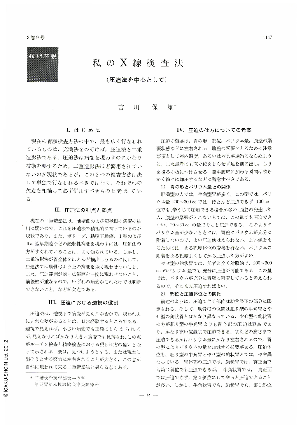
Ⅰ.はじめに
現在の胃腸検査方法の中で,最も広く行なわれているものは,充満法をのぞけば,圧迫法と二重造影法である.圧迫法は病変を現わすのにかなり技術を要するため,二重造影法ほど繁用されていないのが現状であるが,この2つの検査方法は決して単独で行なわれるべきではなく,それぞれの欠点を相補って必ず併用すべきものと考えている.
In recent roentgenological examination of the stomach, compression method has not been used so often as double contrast method. But these two methods should be used together. It is very effective to use compression method for elevated and protruded lesions, but it is not always valid for depressed and excavated lesions. Double contrast method is more effective for this kind of lesion. This difference depends on the visibility of the lesion at the time of fluoroscopy. Degree of difficulty for the compression method depends on the shape of the stomach, site of lesion, volume of barium, and the tension of abdominal wall.
This also depends on the sort of gastric lesion. The change can be imagined from the manifestation of compression image. It is necessary to follow the certain order and to make some diagnosis when X-ray examination is being performed. All parts of the stomach should be taken in case of routine examination. Even in the difficult cases, X-ray photographs by which approximate diagnosis can be made, should be taken. In case of the routine examination, the site of change should be examined rather precisely and many photograhs should be taken. If not, the diagnosis could not be decided afterwards sometimes. X-ray examination should be performed pertinently and the time of fluoroscopy should be made as short as possible. Emphasis should be put on interpretation of films rather than fluoroscopic diagnosis. For this purpose, it is necessary to learn the expedient method and the knack. For the close or the serial examination, good result will be obtained if the method by which clear figure was obtained at the time of routine examination, is used.
Copyright © 1968, Igaku-Shoin Ltd. All rights reserved.